 Movie
Movie Controller

[English] 日本語
 Yorodumi
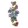
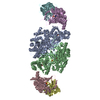
- EMDB-0234: Structure of Influenza Hemagglutinin ectodomain (A/duck/Alberta/35/76) -
+ Open data

- Basic information
| Entry | Database: EMDB / ID: EMD-0234 | ||||||||||||||||||||||||
|---|---|---|---|---|---|---|---|---|---|---|---|---|---|---|---|---|---|---|---|---|---|---|---|---|---|
| Title | Structure of Influenza Hemagglutinin ectodomain (A/duck/Alberta/35/76) | ||||||||||||||||||||||||
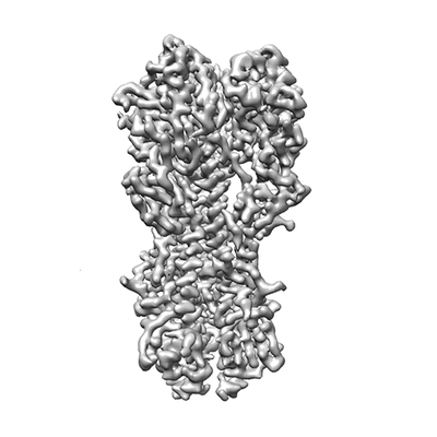
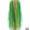
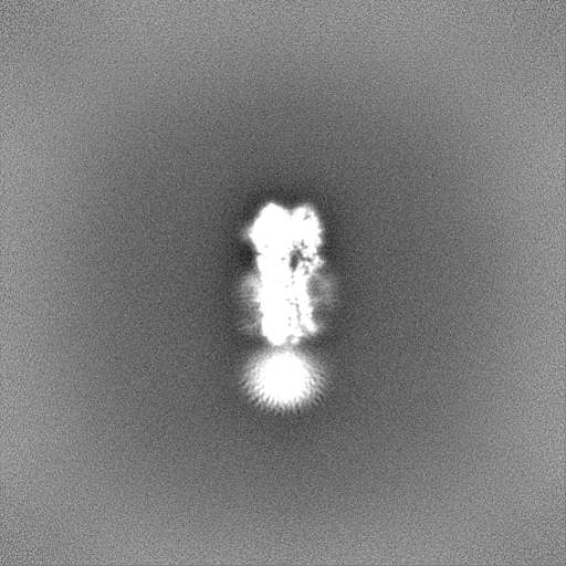
 Map data Map data | Sharpened map of A/duck/Alberta/35/76 Haemagglutinin Ectodomain | ||||||||||||||||||||||||
 Sample Sample |
| ||||||||||||||||||||||||
 Keywords Keywords | Influenza virus / Hemagglutinin / Membrane protein / Membrane fusion / VIRAL PROTEIN | ||||||||||||||||||||||||
| Function / homology |  Function and homology information Function and homology informationviral budding from plasma membrane / clathrin-dependent endocytosis of virus by host cell / host cell surface receptor binding / fusion of virus membrane with host plasma membrane / fusion of virus membrane with host endosome membrane / viral envelope / virion attachment to host cell / host cell plasma membrane / virion membrane / membrane Similarity search - Function | ||||||||||||||||||||||||
| Biological species |  Influenza A virus (strain A/Duck/Alberta/35/1976 H1N1) Influenza A virus (strain A/Duck/Alberta/35/1976 H1N1) | ||||||||||||||||||||||||
| Method | single particle reconstruction / cryo EM / Resolution: 3.3 Å | ||||||||||||||||||||||||
 Authors Authors | Benton DJ / Rosenthal PB | ||||||||||||||||||||||||
| Funding support |  United Kingdom, 7 items United Kingdom, 7 items
| ||||||||||||||||||||||||
 Citation Citation | Journal: Proc Natl Acad Sci U S A / Year: 2018 Title: Influenza hemagglutinin membrane anchor. Authors: Donald J Benton / Andrea Nans / Lesley J Calder / Jack Turner / Ursula Neu / Yi Pu Lin / Esther Ketelaars / Nicole L Kallewaard / Davide Corti / Antonio Lanzavecchia / Steven J Gamblin / ...Authors: Donald J Benton / Andrea Nans / Lesley J Calder / Jack Turner / Ursula Neu / Yi Pu Lin / Esther Ketelaars / Nicole L Kallewaard / Davide Corti / Antonio Lanzavecchia / Steven J Gamblin / Peter B Rosenthal / John J Skehel /   Abstract: Viruses with membranes fuse them with cellular membranes, to transfer their genomes into cells at the beginning of infection. For Influenza virus, the membrane glycoprotein involved in fusion is the ...Viruses with membranes fuse them with cellular membranes, to transfer their genomes into cells at the beginning of infection. For Influenza virus, the membrane glycoprotein involved in fusion is the hemagglutinin (HA), the 3D structure of which is known from X-ray crystallographic studies. The soluble ectodomain fragments used in these studies lacked the "membrane anchor" portion of the molecule. Since this region has a role in membrane fusion, we have determined its structure by analyzing the intact, full-length molecule in a detergent micelle, using cryo-EM. We have also compared the structures of full-length HA-detergent micelles with full-length HA-Fab complex detergent micelles, to describe an infectivity-neutralizing monoclonal Fab that binds near the ectodomain membrane anchor junction. We determine a high-resolution HA structure which compares favorably in detail with the structure of the ectodomain seen by X-ray crystallography; we detect, clearly, all five carbohydrate side chains of HA; and we find that the ectodomain is joined to the membrane anchor by flexible, eight-residue-long, linkers. The linkers extend into the detergent micelle to join a central triple-helical structure that is a major component of the membrane anchor. | ||||||||||||||||||||||||
| History |
|
- Structure visualization
| Movie |
Movie viewer |
|---|---|
| Structure viewer | EM map: SurfViewMolmilJmol/JSmol |
| Supplemental images |
- Downloads & links
-EMDB archive
| Map data | emd_0234.map.gz | 475.6 MB | EMDB map data format | |
|---|---|---|---|---|
| Header (meta data) | emd-0234-v30.xmlemd-0234.xml | 21 KB 21 KB | Display Display | EMDB header |
| FSC (resolution estimation) | emd_0234_fsc.xml | 18.1 KB | Display | FSC data file |
| Images |  emd_0234.png emd_0234.png | 44.2 KB | ||
| Masks | emd_0234_msk_1.map | 512 MB | Mask map | |
| Filedesc metadata | emd-0234.cif.gz | 6.4 KB | ||
| Others | emd_0234_additional.map.gzemd_0234_half_map_1.map.gzemd_0234_half_map_2.map.gz | 475.1 MB 475.4 MB 475.4 MB | ||
| Archive directory |  http://ftp.pdbj.org/pub/emdb/structures/EMD-0234ftp://ftp.pdbj.org/pub/emdb/structures/EMD-0234 http://ftp.pdbj.org/pub/emdb/structures/EMD-0234ftp://ftp.pdbj.org/pub/emdb/structures/EMD-0234 | HTTPS FTP |
-Related structure data
| Related structure data | 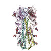 6hjnMC 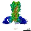 0235C 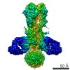 0236C 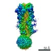 0237C 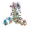 6hjpC 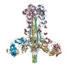 6hjqC  6hjrC 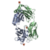 6hkgC M: atomic model generated by this map C: citing same article ( |
|---|---|
| Similar structure data |


-Links
| EMDB pages | EMDB (EBI/PDBe) / EMDataResource |
|---|---|
| Related items in Molecule of the Month |


-Map
| File | Download / File: emd_0234.map.gz / Format: CCP4 / Size: 512 MB / Type: IMAGE STORED AS FLOATING POINT NUMBER (4 BYTES) | ||||||||||||||||||||||||||||||||||||||||||||||||||||||||||||
|---|---|---|---|---|---|---|---|---|---|---|---|---|---|---|---|---|---|---|---|---|---|---|---|---|---|---|---|---|---|---|---|---|---|---|---|---|---|---|---|---|---|---|---|---|---|---|---|---|---|---|---|---|---|---|---|---|---|---|---|---|---|
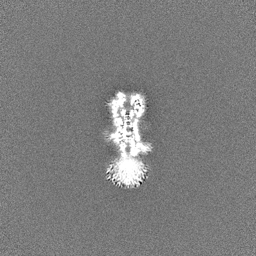
| Annotation | Sharpened map of A/duck/Alberta/35/76 Haemagglutinin Ectodomain | ||||||||||||||||||||||||||||||||||||||||||||||||||||||||||||
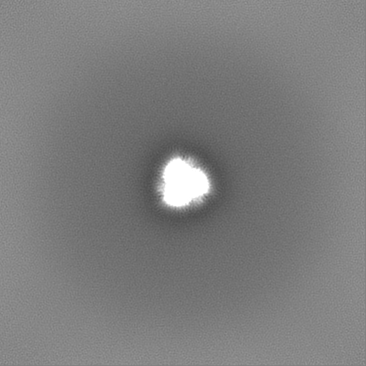
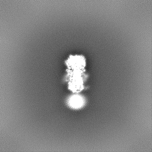
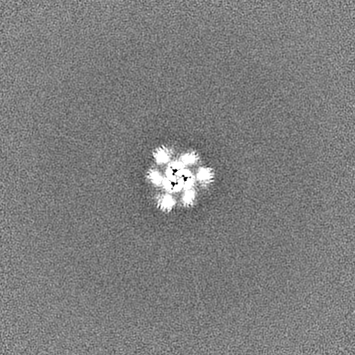
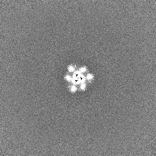
| Projections & slices | Image control
Images are generated by Spider. | ||||||||||||||||||||||||||||||||||||||||||||||||||||||||||||
| Voxel size | X=Y=Z: 1.078 Å | ||||||||||||||||||||||||||||||||||||||||||||||||||||||||||||
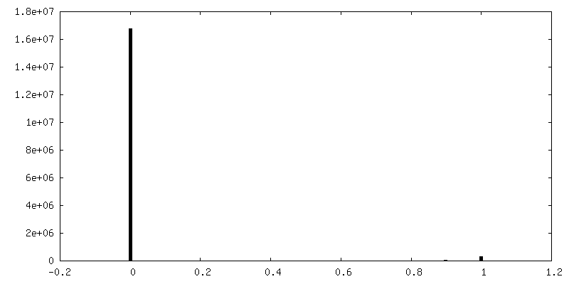
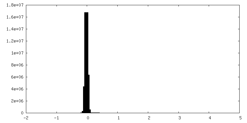
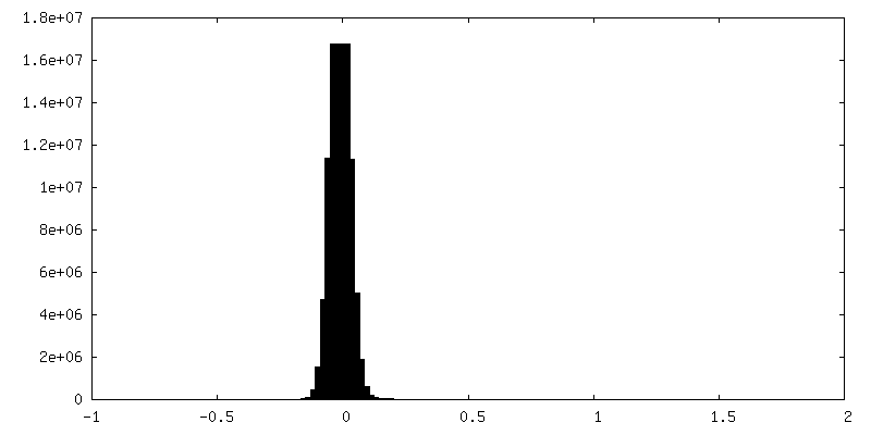
| Density |
| ||||||||||||||||||||||||||||||||||||||||||||||||||||||||||||
| Symmetry | Space group: 1 | ||||||||||||||||||||||||||||||||||||||||||||||||||||||||||||
| Details | EMDB XML:
CCP4 map header:
| ||||||||||||||||||||||||||||||||||||||||||||||||||||||||||||
 Z (Sec.)
 Y (Row.)
 X (Col.)


-Supplemental data
-Mask #1
| File | emd_0234_msk_1.map | ||||||||||||
|---|---|---|---|---|---|---|---|---|---|---|---|---|---|
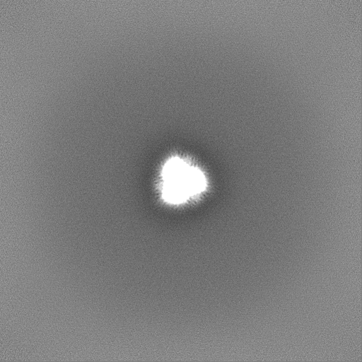
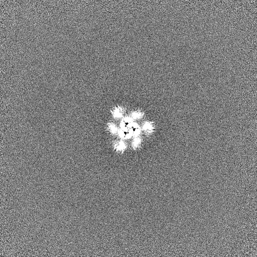
| Projections & Slices |
| ||||||||||||
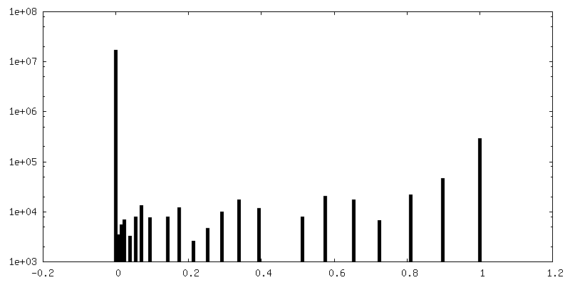
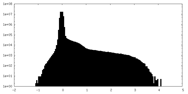
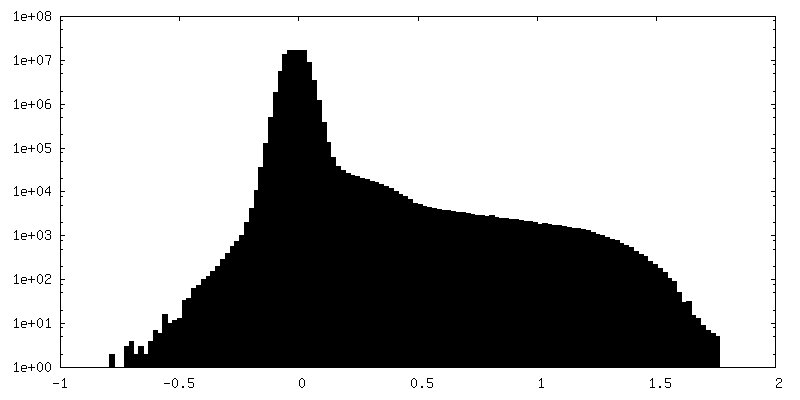
| Density Histograms |


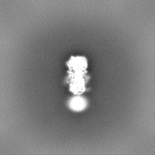
-Additional map: Unsharpened map of A/duck/Alberta/35/76 Hemagglutinin Ectodomain
| File | emd_0234_additional.map | ||||||||||||
|---|---|---|---|---|---|---|---|---|---|---|---|---|---|
| Annotation | Unsharpened map of A/duck/Alberta/35/76 Hemagglutinin Ectodomain | ||||||||||||
| Projections & Slices |
| ||||||||||||
| Density Histograms |


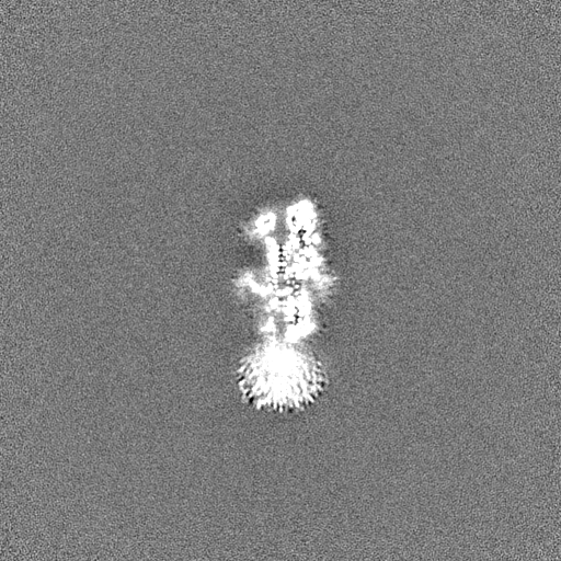
-Half map: Half Map 1 of A/duck/Alberta/35/76 Hemagglutinin Ectodomain
| File | emd_0234_half_map_1.map | ||||||||||||
|---|---|---|---|---|---|---|---|---|---|---|---|---|---|
| Annotation | Half Map 1 of A/duck/Alberta/35/76 Hemagglutinin Ectodomain | ||||||||||||
| Projections & Slices |
| ||||||||||||
| Density Histograms |

-Half map: Half Map 2 of A/duck/Alberta/35/76 Hemagglutinin Ectodomain
| File | emd_0234_half_map_2.map | ||||||||||||
|---|---|---|---|---|---|---|---|---|---|---|---|---|---|
| Annotation | Half Map 2 of A/duck/Alberta/35/76 Hemagglutinin Ectodomain | ||||||||||||
| Projections & Slices |
| ||||||||||||
| Density Histograms |


- Sample components
-Entire : A/duck/Alberta/35/76 Hemagglutinin Ectodomain
| Entire | Name: A/duck/Alberta/35/76 Hemagglutinin Ectodomain |
|---|---|
| Components |
|
-Supramolecule #1: A/duck/Alberta/35/76 Hemagglutinin Ectodomain
| Supramolecule | Name: A/duck/Alberta/35/76 Hemagglutinin Ectodomain / type: complex / ID: 1 / Parent: 0 / Macromolecule list: #1-#2 |
|---|---|
| Source (natural) | Organism: Influenza A virus (strain A/Duck/Alberta/35/1976 H1N1) |
| Molecular weight | Theoretical: 170 KDa |
-Macromolecule #1: Hemagglutinin
| Macromolecule | Name: Hemagglutinin / type: protein_or_peptide / ID: 1 / Number of copies: 3 / Enantiomer: LEVO |
|---|---|
| Source (natural) | Organism: Influenza A virus (strain A/Duck/Alberta/35/1976 H1N1) |
| Molecular weight | Theoretical: 35.571695 KDa |
| Sequence | String: DTICVGYHAN NSTDTVDTVL EKNVTVTHSV NLLEDSHNGK LCSLNGIAPL QLGKCNVAGW LLGNPECDLL LTANSWSYII ETSNSENGT CYPGEFIDYE ELREQLSSVS SFEKFEIFPK ASSWPNHETT KGVTAACSYS GASSFYRNLL WITKKGTSYP K LSKSYTNN ...String: DTICVGYHAN NSTDTVDTVL EKNVTVTHSV NLLEDSHNGK LCSLNGIAPL QLGKCNVAGW LLGNPECDLL LTANSWSYII ETSNSENGT CYPGEFIDYE ELREQLSSVS SFEKFEIFPK ASSWPNHETT KGVTAACSYS GASSFYRNLL WITKKGTSYP K LSKSYTNN KGKEVLVLWG VHHPPSVSEQ QSLYQNADAY VSVGSSKYNR RFAPEIAARP KVRGQAGRMN YYWTLLDQGD TI TFEATGN LIAPWYAFAL NKGSDSGIIT SDAPVHNCDT RCQTPHGALN SSLPFQNVHP ITIGECPKYV KSTKLRMATG LRN VPS UniProtKB: Hemagglutinin |
-Macromolecule #2: Hemagglutinin
| Macromolecule | Name: Hemagglutinin / type: protein_or_peptide / ID: 2 / Number of copies: 3 / Enantiomer: LEVO |
|---|---|
| Source (natural) | Organism: Influenza A virus (strain A/Duck/Alberta/35/1976 H1N1) |
| Molecular weight | Theoretical: 19.838834 KDa |
| Sequence | String: GLFGAIAGFI EGGWTGMIDG WYGYHHQNEQ GSGYAADQKS TQNAIDGITS KVNSVIEKMN TQFTAVGKEF NNLERRIENL NKKVDDGFL DVWTYNAELL VLLENERTLD FHDSNVRNLY EKVKSQLRNN AKEIGNGCFE FYHKCDDECM ESVKNGTYDY P KYSEESKL NREEI UniProtKB: Hemagglutinin |
-Macromolecule #7: UNKNOWN BRANCHED FRAGMENT OF PHOSPHOLIPID
| Macromolecule | Name: UNKNOWN BRANCHED FRAGMENT OF PHOSPHOLIPID / type: ligand / ID: 7 / Number of copies: 3 / Formula: UPL |
|---|---|
| Molecular weight | Theoretical: 478.92 Da |
| Chemical component information | 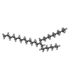 ChemComp-UPL: |
-Experimental details
-Structure determination
| Method | cryo EM |
|---|---|
 Processing Processing | single particle reconstruction |
| Aggregation state | particle |
-Sample preparation
| Concentration | 2 mg/mL | ||||||||||||||
|---|---|---|---|---|---|---|---|---|---|---|---|---|---|---|---|
| Buffer | pH: 8 Component:
| ||||||||||||||
| Vitrification | Cryogen name: ETHANE / Instrument: FEI VITROBOT MARK III |
- Electron microscopy
| Microscope | FEI TITAN KRIOS |
|---|---|
| Specialist optics | Energy filter - Name: GIF Quantum LS / Energy filter - Slit width: 20 eV |
| Image recording | Film or detector model: GATAN K2 SUMMIT (4k x 4k) / Detector mode: COUNTING / Average electron dose: 43.0 e/Å2 |
| Electron beam | Acceleration voltage: 300 kV / Electron source:  FIELD EMISSION GUN FIELD EMISSION GUN |
| Electron optics | C2 aperture diameter: 50.0 µm / Illumination mode: FLOOD BEAM / Imaging mode: BRIGHT FIELD / Cs: 2.7 mm |
| Sample stage | Specimen holder model: FEI TITAN KRIOS AUTOGRID HOLDER / Cooling holder cryogen: NITROGEN |
| Experimental equipment |  Model: Titan Krios / Image courtesy: FEI Company |

