 Movie
Movie Controller

[English] 日本語
 Yorodumi
- EMDB-0165: Immature CA domain from HIV-1 MA-CA Gag proteolytic cleavage muta... -
+ Open data

- Basic information
| Entry | Database: EMDB / ID: EMD-0165 | |||||||||
|---|---|---|---|---|---|---|---|---|---|---|
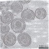
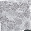
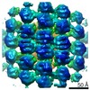
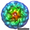
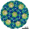
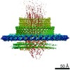
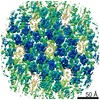
| Title | Immature CA domain from HIV-1 MA-CA Gag proteolytic cleavage mutant virus particles | |||||||||
 Map data Map data | Structure of the immature CA domain from HIV-1 MA-CA Gag proteolytic cleavage mutant virus particles | |||||||||
 Sample Sample |
| |||||||||
| Biological species |  Homo sapiens (human) / Homo sapiens (human) /   Human immunodeficiency virus 1 Human immunodeficiency virus 1 | |||||||||
| Method | subtomogram averaging / cryo EM / Resolution: 3.7 Å | |||||||||
 Authors Authors | Mattei S / Tan AWK / Glass B / Mueller B / Kraeusslich HG / Briggs JAG | |||||||||
 Citation Citation | Journal: Proc Natl Acad Sci U S A / Year: 2018 Title: High-resolution structures of HIV-1 Gag cleavage mutants determine structural switch for virus maturation. Authors: Simone Mattei / Aaron Tan / Bärbel Glass / Barbara Müller / Hans-Georg Kräusslich / John A G Briggs /    Abstract: HIV-1 maturation occurs via multiple proteolytic cleavages of the Gag polyprotein, causing rearrangement of the virus particle required for infectivity. Cleavage results in beta-hairpin formation at ...HIV-1 maturation occurs via multiple proteolytic cleavages of the Gag polyprotein, causing rearrangement of the virus particle required for infectivity. Cleavage results in beta-hairpin formation at the N terminus of the CA (capsid) protein and loss of a six-helix bundle formed by the C terminus of CA and the neighboring SP1 peptide. How individual cleavages contribute to changes in protein structure and interactions, and how the mature, conical capsid forms, are poorly understood. Here, we employed cryoelectron tomography to determine morphology and high-resolution CA lattice structures for HIV-1 derivatives in which Gag cleavage sites are mutated. These analyses prompt us to revise current models for the crucial maturation switch. Unlike previously proposed, cleavage on either terminus of CA was sufficient, in principle, for lattice maturation, while complete processing was needed for conical capsid formation. We conclude that destabilization of the six-helix bundle, rather than beta-hairpin formation, represents the main determinant of structural maturation. | |||||||||
| History |
|
- Structure visualization
| Movie |
 Movie viewer Movie viewer |
|---|---|
| Structure viewer | EM map: SurfViewMolmilJmol/JSmol |
| Supplemental images |

- Downloads & links
-EMDB archive
| Map data | emd_0165.map.gz | 25 MB | EMDB map data format | |
|---|---|---|---|---|
| Header (meta data) | emd-0165-v30.xmlemd-0165.xml | 13.9 KB 13.9 KB | Display Display | EMDB header |
| Images | 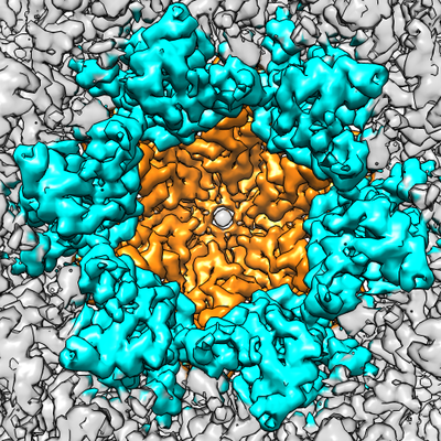 emd_0165.png emd_0165.png | 321.2 KB | ||
| Archive directory |  http://ftp.pdbj.org/pub/emdb/structures/EMD-0165ftp://ftp.pdbj.org/pub/emdb/structures/EMD-0165 http://ftp.pdbj.org/pub/emdb/structures/EMD-0165ftp://ftp.pdbj.org/pub/emdb/structures/EMD-0165 | HTTPS FTP |
-Related structure data
| Related structure data |  0164C  0166C  0167C 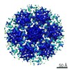 0168C 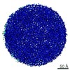 0169C 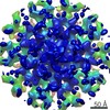 0170C 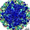 0171C C: citing same article ( |
|---|---|
| Similar structure data |

-Links
| EMDB pages | EMDB (EBI/PDBe) / EMDataResource |
|---|---|
| Related items in Molecule of the Month |

-Map
| File | Download / File: emd_0165.map.gz / Format: CCP4 / Size: 27 MB / Type: IMAGE STORED AS FLOATING POINT NUMBER (4 BYTES) | ||||||||||||||||||||||||||||||||||||||||||||||||||||||||||||
|---|---|---|---|---|---|---|---|---|---|---|---|---|---|---|---|---|---|---|---|---|---|---|---|---|---|---|---|---|---|---|---|---|---|---|---|---|---|---|---|---|---|---|---|---|---|---|---|---|---|---|---|---|---|---|---|---|---|---|---|---|---|
| Annotation | Structure of the immature CA domain from HIV-1 MA-CA Gag proteolytic cleavage mutant virus particles | ||||||||||||||||||||||||||||||||||||||||||||||||||||||||||||
| Projections & slices | Image control
Images are generated by Spider. | ||||||||||||||||||||||||||||||||||||||||||||||||||||||||||||
| Voxel size | X=Y=Z: 1.35 Å | ||||||||||||||||||||||||||||||||||||||||||||||||||||||||||||
| Density |
| ||||||||||||||||||||||||||||||||||||||||||||||||||||||||||||
| Symmetry | Space group: 1 | ||||||||||||||||||||||||||||||||||||||||||||||||||||||||||||
| Details | EMDB XML:
CCP4 map header:
| ||||||||||||||||||||||||||||||||||||||||||||||||||||||||||||
 Z (Sec.)
 Y (Row.)
 X (Col.)


-Supplemental data
- Sample components
-Entire : Human immunodeficiency virus 1
| Entire | Name: Human immunodeficiency virus 1 |
|---|---|
| Components |
|
-Supramolecule #1: Human immunodeficiency virus 1
| Supramolecule | Name: Human immunodeficiency virus 1 / type: virus / ID: 1 / Parent: 0 / Macromolecule list: all / NCBI-ID: 11676 / Sci species name: Human immunodeficiency virus 1 / Virus type: VIRION / Virus isolate: STRAIN / Virus enveloped: Yes / Virus empty: No |
|---|---|
| Host (natural) | Organism: Homo sapiens (human) |
| Host system | Organism: Homo sapiens (human) / Recombinant cell: HEK293T / Recombinant plasmid: pCHIV |
-Macromolecule #1: Gag polyprotein
| Macromolecule | Name: Gag polyprotein / type: protein_or_peptide / ID: 1 / Enantiomer: LEVO |
|---|---|
| Source (natural) | Organism: Homo sapiens (human) |
| Sequence | String: MGARASVLSG GELDRWEKIR LRPGGKKKYK LKHIVWASRE LERFAVNPGL LETSEGCRQI LGQLQPSLQ TGSEELRSLY NTVATLYCVH QRIEIKDTKE ALDKIEEEQN KSKKKAQQAA A DTGHSNQV SQNIPIVQNI QGQMVHQAIS PRTLNAWVKV VEEKAFSPEV ...String: MGARASVLSG GELDRWEKIR LRPGGKKKYK LKHIVWASRE LERFAVNPGL LETSEGCRQI LGQLQPSLQ TGSEELRSLY NTVATLYCVH QRIEIKDTKE ALDKIEEEQN KSKKKAQQAA A DTGHSNQV SQNIPIVQNI QGQMVHQAIS PRTLNAWVKV VEEKAFSPEV IPMFSALSEG AT PQDLNTM LNTVGGHQAA MQMLKETINE EAAEWDRVHP VHAGPIAPGQ MREPRGSDIA GTT STLQEQ IGWMTNNPPI PVGEIYKRWI ILGLNKIVRM YSPTSILDIR QGPKEPFRDY VDRF YKTLR AEQASQEVKN WMTETLLVQN ANPDCKTILK ALGPAATLEE MMTACQGVGG PGHKA RVLA EAMSQVTNSA TIMMQRGNFR NQRKIVKCFN CGKEGHTARN CRAPRKKGCW KCGKEG HQM KDCTERQANF LGKIWPSYKG RPGNFLQSRP EPTAPPEESF RSGVETTTPP QKQEPID KE LYPLTSLRSL FGNDPSSQ |
-Experimental details
-Structure determination
| Method | cryo EM |
|---|---|
 Processing Processing | subtomogram averaging |
| Aggregation state | particle |
-Sample preparation
| Buffer | pH: 7.4 |
|---|---|
| Grid | Model: C-flat-2/2 / Material: COPPER / Mesh: 300 / Support film - Material: CARBON / Support film - topology: HOLEY / Pretreatment - Type: GLOW DISCHARGE / Pretreatment - Atmosphere: AIR Details: The grid was glow discharged with a current of 20 mA |
| Vitrification | Cryogen name: ETHANE / Chamber humidity: 100 % / Chamber temperature: 288.15 K / Instrument: FEI VITROBOT MARK II |
- Electron microscopy
| Microscope | FEI TITAN KRIOS |
|---|---|
| Specialist optics | Energy filter - Name: GIF Quantum LS / Energy filter - Slit width: 20 eV |
| Image recording | Film or detector model: GATAN K2 QUANTUM (4k x 4k) / Detector mode: SUPER-RESOLUTION / Digitization - Frames/image: 1-21 / Average electron dose: 3.7 e/Å2 |
| Electron beam | Acceleration voltage: 300 kV / Electron source:  FIELD EMISSION GUN FIELD EMISSION GUN |
| Electron optics | C2 aperture diameter: 70.0 µm / Illumination mode: FLOOD BEAM / Imaging mode: BRIGHT FIELD / Cs: 2.7 mm / Nominal defocus max: 5.0 µm / Nominal defocus min: 1.5 µm |
| Sample stage | Specimen holder model: FEI TITAN KRIOS AUTOGRID HOLDER / Cooling holder cryogen: NITROGEN |
| Experimental equipment |  Model: Titan Krios / Image courtesy: FEI Company |
