Movie
Movie Controller

[English] 日本語
 Yorodumi
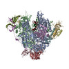
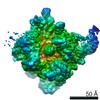
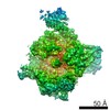
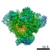
- EMDB-0147: Yeast RNA polymerase I elongation complex stalled by cyclobutane ... -
+ Open data

- Basic information
| Entry | Database: EMDB / ID: EMD-0147 | |||||||||
|---|---|---|---|---|---|---|---|---|---|---|
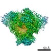
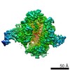
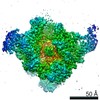
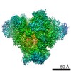
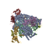
| Title | Yeast RNA polymerase I elongation complex stalled by cyclobutane pyrimidine dimer (CPD) with fully-ordered A49 | |||||||||
 Map data Map data | ||||||||||
 Sample Sample |
| |||||||||
 Keywords Keywords | RNA Polymerase I / UV-damage / stalling / Cyclobutane Pyrimidine Dimers (CPD) / TRANSCRIPTION | |||||||||
| Function / homology |  Function and homology information Function and homology informationnuclear DNA-directed RNA polymerase complex / RNA polymerase I preinitiation complex assembly / RNA Polymerase I Transcription Initiation / Processing of Capped Intron-Containing Pre-mRNA / RNA Polymerase III Transcription Initiation From Type 2 Promoter / regulation of cell size / RNA Pol II CTD phosphorylation and interaction with CE / Formation of the Early Elongation Complex / mRNA Capping / RNA polymerase II transcribes snRNA genes ...nuclear DNA-directed RNA polymerase complex / RNA polymerase I preinitiation complex assembly / RNA Polymerase I Transcription Initiation / Processing of Capped Intron-Containing Pre-mRNA / RNA Polymerase III Transcription Initiation From Type 2 Promoter / regulation of cell size / RNA Pol II CTD phosphorylation and interaction with CE / Formation of the Early Elongation Complex / mRNA Capping / RNA polymerase II transcribes snRNA genes / TP53 Regulates Transcription of DNA Repair Genes / RNA Polymerase II Promoter Escape / RNA Polymerase II Transcription Pre-Initiation And Promoter Opening / RNA Polymerase II Transcription Initiation / RNA Polymerase II Transcription Initiation And Promoter Clearance / RNA Polymerase II Pre-transcription Events / termination of RNA polymerase III transcription / Formation of TC-NER Pre-Incision Complex / RNA-templated transcription / RNA Polymerase I Promoter Escape / termination of RNA polymerase I transcription / transcription initiation at RNA polymerase III promoter / Gap-filling DNA repair synthesis and ligation in TC-NER / nucleolar large rRNA transcription by RNA polymerase I / transcription initiation at RNA polymerase I promoter / Estrogen-dependent gene expression / transcription by RNA polymerase III / Dual incision in TC-NER / RNA polymerase I complex / RNA polymerase III complex / transcription elongation by RNA polymerase I / RNA polymerase II, core complex / tRNA transcription by RNA polymerase III / transcription by RNA polymerase I / transcription initiation at RNA polymerase II promoter / promoter-specific chromatin binding / transcription elongation by RNA polymerase II / ribonucleoside binding / DNA-directed RNA polymerase / DNA-directed RNA polymerase activity / peroxisome / ribosome biogenesis / transcription by RNA polymerase II / nucleic acid binding / RNA polymerase II-specific DNA-binding transcription factor binding / protein dimerization activity / nucleolus / negative regulation of transcription by RNA polymerase II / DNA binding / zinc ion binding / nucleoplasm / metal ion binding / nucleus / cytoplasm Similarity search - Function | |||||||||
| Biological species |  | |||||||||
| Method | single particle reconstruction / cryo EM / Resolution: 4.6 Å | |||||||||
 Authors Authors | Sanz-Murillo M / Xu J | |||||||||
| Funding support |  Spain, 2 items Spain, 2 items
| |||||||||
 Citation Citation | Journal: Proc Natl Acad Sci U S A / Year: 2018 Title: Structural basis of RNA polymerase I stalling at UV light-induced DNA damage. Authors: Marta Sanz-Murillo / Jun Xu / Georgiy A Belogurov / Olga Calvo / David Gil-Carton / María Moreno-Morcillo / Dong Wang / Carlos Fernández-Tornero /   Abstract: RNA polymerase I (Pol I) transcribes ribosomal DNA (rDNA) to produce the ribosomal RNA (rRNA) precursor, which accounts for up to 60% of the total transcriptional activity in growing cells. Pol I ...RNA polymerase I (Pol I) transcribes ribosomal DNA (rDNA) to produce the ribosomal RNA (rRNA) precursor, which accounts for up to 60% of the total transcriptional activity in growing cells. Pol I monitors rDNA integrity and influences cell survival, but little is known about how this enzyme processes UV-induced lesions. We report the electron cryomicroscopy structure of Pol I in an elongation complex containing a cyclobutane pyrimidine dimer (CPD) at a resolution of 3.6 Å. The structure shows that the lesion induces an early translocation intermediate exhibiting unique features. The bridge helix residue Arg1015 plays a major role in CPD-induced Pol I stalling, as confirmed by mutational analysis. These results, together with biochemical data presented here, reveal the molecular mechanism of Pol I stalling by CPD lesions, which is distinct from Pol II arrest by CPD lesions. Our findings open the avenue to unravel the molecular mechanisms underlying cell endurance to lesions on rDNA. | |||||||||
| History |
|
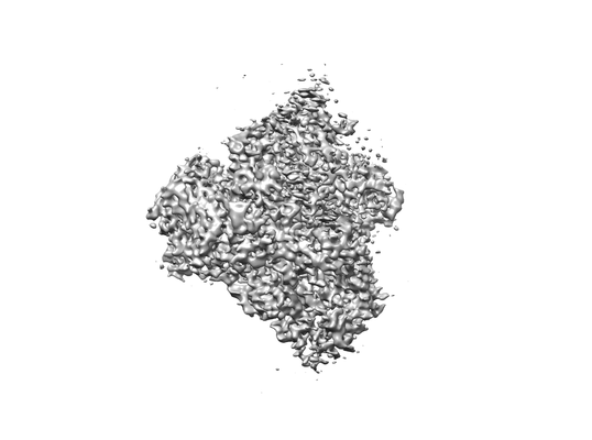
- Structure visualization
| Movie |
Movie viewer |
|---|---|
| Structure viewer | EM map: SurfViewMolmilJmol/JSmol |
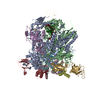
| Supplemental images |
- Downloads & links
-EMDB archive
| Map data | emd_0147.map.gz | 85.3 MB | EMDB map data format | |
|---|---|---|---|---|
| Header (meta data) | emd-0147-v30.xmlemd-0147.xml | 36.2 KB 36.2 KB | Display Display | EMDB header |
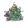
| Images |  emd_0147.png emd_0147.png | 38.1 KB | ||
| Filedesc metadata | emd-0147.cif.gz | 10.6 KB | ||
| Archive directory |  http://ftp.pdbj.org/pub/emdb/structures/EMD-0147ftp://ftp.pdbj.org/pub/emdb/structures/EMD-0147 http://ftp.pdbj.org/pub/emdb/structures/EMD-0147ftp://ftp.pdbj.org/pub/emdb/structures/EMD-0147 | HTTPS FTP |
-Related structure data
| Related structure data | 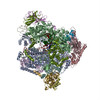 6h68MC 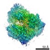 0146C 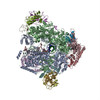 6h67C M: atomic model generated by this map C: citing same article ( |
|---|---|
| Similar structure data |
-Links
| EMDB pages | EMDB (EBI/PDBe) / EMDataResource |
|---|---|
| Related items in Molecule of the Month |


-Map
| File | Download / File: emd_0147.map.gz / Format: CCP4 / Size: 91.1 MB / Type: IMAGE STORED AS FLOATING POINT NUMBER (4 BYTES) | ||||||||||||||||||||||||||||||||||||||||||||||||||||||||||||
|---|---|---|---|---|---|---|---|---|---|---|---|---|---|---|---|---|---|---|---|---|---|---|---|---|---|---|---|---|---|---|---|---|---|---|---|---|---|---|---|---|---|---|---|---|---|---|---|---|---|---|---|---|---|---|---|---|---|---|---|---|---|
| Projections & slices | Image control
Images are generated by Spider. | ||||||||||||||||||||||||||||||||||||||||||||||||||||||||||||
| Voxel size | X=Y=Z: 1.06 Å | ||||||||||||||||||||||||||||||||||||||||||||||||||||||||||||
| Density |
| ||||||||||||||||||||||||||||||||||||||||||||||||||||||||||||
| Symmetry | Space group: 1 | ||||||||||||||||||||||||||||||||||||||||||||||||||||||||||||
| Details | EMDB XML:
CCP4 map header:
| ||||||||||||||||||||||||||||||||||||||||||||||||||||||||||||
 Z (Sec.)
 Y (Row.)
 X (Col.)


-Supplemental data
- Sample components
+Entire : RNA Polymerase I stalling at DNA damage
+Supramolecule #1: RNA Polymerase I stalling at DNA damage
+Supramolecule #2: RNA Polymerase I stalling at DNA damage
+Supramolecule #3: RNA Polymerase I stalling at DNA damage
+Macromolecule #1: DNA-directed RNA polymerase I subunit RPA190
+Macromolecule #2: DNA-directed RNA polymerase I subunit RPA135
+Macromolecule #3: DNA-directed RNA polymerases I and III subunit RPAC1
+Macromolecule #4: DNA-directed RNA polymerase I subunit RPA14
+Macromolecule #5: DNA-directed RNA polymerases I, II, and III subunit RPABC1
+Macromolecule #6: DNA-directed RNA polymerases I, II, and III subunit RPABC2
+Macromolecule #7: DNA-directed RNA polymerase I subunit RPA43
+Macromolecule #8: DNA-directed RNA polymerases I, II, and III subunit RPABC3
+Macromolecule #9: DNA-directed RNA polymerase I subunit RPA12
+Macromolecule #10: DNA-directed RNA polymerases I, II, and III subunit RPABC5
+Macromolecule #11: DNA-directed RNA polymerases I and III subunit RPAC2
+Macromolecule #12: DNA-directed RNA polymerases I, II, and III subunit RPABC4
+Macromolecule #13: DNA-directed RNA polymerase I subunit RPA49
+Macromolecule #14: DNA-directed RNA polymerase I subunit RPA34
+Macromolecule #15: RNA
+Macromolecule #16: Template DNA
+Macromolecule #17: Non-template DNA
+Macromolecule #18: ZINC ION
-Experimental details
-Structure determination
| Method | cryo EM |
|---|---|
 Processing Processing | single particle reconstruction |
| Aggregation state | particle |
-Sample preparation
| Concentration | 0.22 mg/mL | ||||||||||||
|---|---|---|---|---|---|---|---|---|---|---|---|---|---|
| Buffer | pH: 8 Component:
| ||||||||||||
| Grid | Model: Quantifoil R2/1 / Material: COPPER | ||||||||||||
| Vitrification | Cryogen name: ETHANE / Chamber humidity: 95 % / Chamber temperature: 277.15 K / Instrument: FEI VITROBOT MARK III |
- Electron microscopy
| Microscope | FEI TITAN KRIOS |
|---|---|
| Image recording | Film or detector model: GATAN K2 SUMMIT (4k x 4k) / Detector mode: COUNTING / Number grids imaged: 1 / Number real images: 2056 / Average electron dose: 5.25 e/Å2 |
| Electron beam | Acceleration voltage: 300 kV / Electron source:  FIELD EMISSION GUN FIELD EMISSION GUN |
| Electron optics | Illumination mode: FLOOD BEAM / Imaging mode: BRIGHT FIELD / Cs: 2.7 mm / Nominal defocus max: 4.0 µm / Nominal defocus min: 1.5 µm |
| Sample stage | Specimen holder model: FEI TITAN KRIOS AUTOGRID HOLDER / Cooling holder cryogen: NITROGEN |
| Experimental equipment |  Model: Titan Krios / Image courtesy: FEI Company |
+Image processing
-Atomic model buiding 1
| Initial model |
| ||||||
|---|---|---|---|---|---|---|---|
| Refinement | Space: REAL / Protocol: AB INITIO MODEL / Overall B value: 93.9 | ||||||
| Output model | PDB-6h68: |