 Movie
Movie Controller

[English] 日本語
 Yorodumi
- EMDB-0117: Subtomogram averaging of nucleosome particles decorating chromati... -
+ Open data

- Basic information
| Entry | Database: EMDB / ID: EMD-0117 | |||||||||
|---|---|---|---|---|---|---|---|---|---|---|
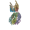
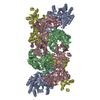
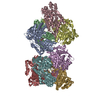
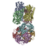
| Title | Subtomogram averaging of nucleosome particles decorating chromatin plates from metaphase chromosomes | |||||||||
 Map data Map data | Structure of decorative particles | |||||||||
 Sample Sample |
| |||||||||
| Biological species |  Homo sapiens (human) Homo sapiens (human) | |||||||||
| Method | subtomogram averaging / cryo EM / Resolution: 30.0 Å | |||||||||
 Authors Authors | Chicano A / Crosas E / Engel BD / Melero R / Oton J / Daban JR | |||||||||
| Funding support |  Spain, 1 items Spain, 1 items
| |||||||||
 Citation Citation | Journal: EMBO J / Year: 2019 Title: Frozen-hydrated chromatin from metaphase chromosomes has an interdigitated multilayer structure. Authors: Andrea Chicano / Eva Crosas / Joaquín Otón / Roberto Melero / Benjamin D Engel / Joan-Ramon Daban /  Abstract: Cryo-electron tomography and small-angle X-ray scattering were used to investigate the chromatin folding in metaphase chromosomes. The tomographic 3D reconstructions show that frozen-hydrated ...Cryo-electron tomography and small-angle X-ray scattering were used to investigate the chromatin folding in metaphase chromosomes. The tomographic 3D reconstructions show that frozen-hydrated chromatin emanated from chromosomes is planar and forms multilayered plates. The layer thickness was measured accounting for the contrast transfer function fringes at the plate edges, yielding a width of ~ 7.5 nm, which is compatible with the dimensions of a monolayer of nucleosomes slightly tilted with respect to the layer surface. Individual nucleosomes are visible decorating distorted plates, but typical plates are very dense and nucleosomes are not identifiable as individual units, indicating that they are tightly packed. Two layers in contact are ~ 13 nm thick, which is thinner than the sum of two independent layers, suggesting that nucleosomes in the layers interdigitate. X-ray scattering of whole chromosomes shows a main scattering peak at ~ 6 nm, which can be correlated with the distance between layers and between interdigitating nucleosomes interacting through their faces. These observations support a model where compact chromosomes are composed of many chromatin layers stacked along the chromosome axis. | |||||||||
| History |
|
- Structure visualization
| Movie |
 Movie viewer Movie viewer |
|---|---|
| Structure viewer | EM map: SurfViewMolmilJmol/JSmol |
| Supplemental images |

- Downloads & links
-EMDB archive
| Map data | emd_0117.map.gz | 449.1 KB | EMDB map data format | |
|---|---|---|---|---|
| Header (meta data) | emd-0117-v30.xmlemd-0117.xml | 9.8 KB 9.8 KB | Display Display | EMDB header |
| Images | 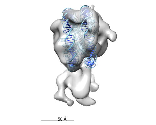 emd_0117.png emd_0117.png | 78.5 KB | ||
| Archive directory |  http://ftp.pdbj.org/pub/emdb/structures/EMD-0117ftp://ftp.pdbj.org/pub/emdb/structures/EMD-0117 http://ftp.pdbj.org/pub/emdb/structures/EMD-0117ftp://ftp.pdbj.org/pub/emdb/structures/EMD-0117 | HTTPS FTP |
-Validation report
| Summary document | emd_0117_validation.pdf.gz | 221.7 KB | Display | EMDB validaton report |
|---|---|---|---|---|
| Full document | emd_0117_full_validation.pdf.gz | 220.8 KB | Display | |
| Data in XML | emd_0117_validation.xml.gz | 4.7 KB | Display | |
| Arichive directory | https://ftp.pdbj.org/pub/emdb/validation_reports/EMD-0117ftp://ftp.pdbj.org/pub/emdb/validation_reports/EMD-0117 | HTTPS FTP |
-Related structure data


-Links
| EMDB pages | EMDB (EBI/PDBe) / EMDataResource |
|---|
-Map
| File | Download / File: emd_0117.map.gz / Format: CCP4 / Size: 489.3 KB / Type: IMAGE STORED AS FLOATING POINT NUMBER (4 BYTES) | ||||||||||||||||||||||||||||||||||||||||||||||||||||||||||||
|---|---|---|---|---|---|---|---|---|---|---|---|---|---|---|---|---|---|---|---|---|---|---|---|---|---|---|---|---|---|---|---|---|---|---|---|---|---|---|---|---|---|---|---|---|---|---|---|---|---|---|---|---|---|---|---|---|---|---|---|---|---|
| Annotation | Structure of decorative particles | ||||||||||||||||||||||||||||||||||||||||||||||||||||||||||||
| Projections & slices | Image control
Images are generated by Spider. | ||||||||||||||||||||||||||||||||||||||||||||||||||||||||||||
| Voxel size | X=Y=Z: 4.21 Å | ||||||||||||||||||||||||||||||||||||||||||||||||||||||||||||
| Density |
| ||||||||||||||||||||||||||||||||||||||||||||||||||||||||||||
| Symmetry | Space group: 1 | ||||||||||||||||||||||||||||||||||||||||||||||||||||||||||||
| Details | EMDB XML:
CCP4 map header:
| ||||||||||||||||||||||||||||||||||||||||||||||||||||||||||||
 Z (Sec.)
 Y (Row.)
 X (Col.)


-Supplemental data
- Sample components
-Entire : Chromatin plates from metaphase chromosomes
| Entire | Name: Chromatin plates from metaphase chromosomes |
|---|---|
| Components |
|
-Supramolecule #1: Chromatin plates from metaphase chromosomes
| Supramolecule | Name: Chromatin plates from metaphase chromosomes / type: complex / ID: 1 / Parent: 0 / Macromolecule list: #1 / Details: Nucleosome particles in the plates |
|---|---|
| Source (natural) | Organism: Homo sapiens (human) / Strain: HeLa / Organelle: Metaphase chromosomes |
-Experimental details
-Structure determination
| Method | cryo EM |
|---|---|
 Processing Processing | subtomogram averaging |
| Aggregation state | particle |
-Sample preparation
| Buffer | pH: 7.2 / Details: 5 mM Pipes (pH 7.2), 5 mM NaCl, and 5 mM MgCl2 |
|---|---|
| Vitrification | Cryogen name: ETHANE-PROPANE / Instrument: FEI VITROBOT MARK III |
| Details | Chromatin emanated from soft-denatured metaphase chromosomes |
- Electron microscopy
| Microscope | FEI TITAN KRIOS |
|---|---|
| Specialist optics | Phase plate: VOLTA PHASE PLATE / Energy filter - Name: GIF 2002 |
| Image recording | #0 - Image recording ID: 1 / #0 - Film or detector model: GATAN K2 SUMMIT (4k x 4k) / #0 - Average electron dose: 1.8 e/Å2 / #1 - Image recording ID: 2 / #1 - Film or detector model: GATAN K2 SUMMIT (4k x 4k) / #1 - Average electron dose: 1.8 e/Å2 |
| Electron beam | Acceleration voltage: 300 kV / Electron source:  FIELD EMISSION GUN FIELD EMISSION GUN |
| Electron optics | Calibrated defocus min: 0.5 µm / Calibrated magnification: 33000 / Illumination mode: OTHER / Imaging mode: BRIGHT FIELD |
| Experimental equipment |  Model: Titan Krios / Image courtesy: FEI Company |
-Image processing
| Image recording ID | 1 |
|---|---|
| Final reconstruction | Number classes used: 4 / Algorithm: FOURIER SPACE / Resolution.type: BY AUTHOR / Resolution: 30.0 Å / Resolution method: OTHER / Details: MonoRes / Number subtomograms used: 315 |
| Extraction | Number tomograms: 1 / Number images used: 902 / Reference model: de novo / Method: manually / Software - Name: EMAN2 |
| Final angle assignment | Type: MAXIMUM LIKELIHOOD / Software - Name: Xmipp (ver. 2.4) |
