 Movie
Movie Controller

[English] 日本語
 Yorodumi
- EMDB-0045: Negative stain EM map of the inner membrane protein GspF (XcpS) o... -
+ Open data

- Basic information
| Entry |  | ||||||||||||
|---|---|---|---|---|---|---|---|---|---|---|---|---|---|
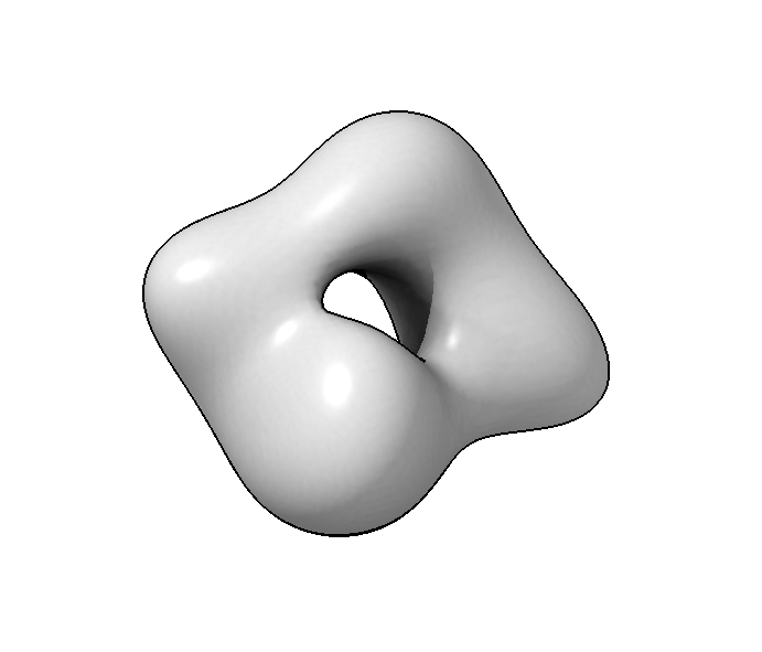
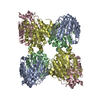
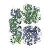
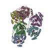
| Title | Negative stain EM map of the inner membrane protein GspF (XcpS) of the bacterial type II secretion system | ||||||||||||
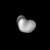
 Map data Map data | Final map after 3D refinement (Relion) low-pass filtered to 2nm. | ||||||||||||
 Sample Sample |
| ||||||||||||
| Biological species |   Pseudomonas aeruginosa (bacteria) Pseudomonas aeruginosa (bacteria) | ||||||||||||
| Method | single particle reconstruction / negative staining / Resolution: 20.0 Å | ||||||||||||
 Authors Authors | Van Putte W / Savvides S | ||||||||||||
| Funding support |  Belgium, Belgium,  Germany, 3 items Germany, 3 items
| ||||||||||||
 Citation Citation | Journal: To Be Published Title: The inner membrane protein GspF of the bacterial type II secretion system adopts a dimeric structure to mediate pseudopilus biogenesis Authors: Van Putte W / Savvides S / Kudryashev M / De Vos T | ||||||||||||
| History |
|
- Structure visualization
| Structure viewer | EM map:  SurfViewMolmilJmol/JSmol SurfViewMolmilJmol/JSmol |
|---|---|
| Supplemental images |
- Downloads & links
-EMDB archive
| Map data | emd_0045.map.gz | 3.5 MB | EMDB map data format | |
|---|---|---|---|---|
| Header (meta data) | emd-0045-v30.xmlemd-0045.xml | 9.2 KB 9.2 KB | Display Display | EMDB header |
| Images |  emd_0045.png emd_0045.png | 82.9 KB | ||
| Archive directory |  http://ftp.pdbj.org/pub/emdb/structures/EMD-0045ftp://ftp.pdbj.org/pub/emdb/structures/EMD-0045 http://ftp.pdbj.org/pub/emdb/structures/EMD-0045ftp://ftp.pdbj.org/pub/emdb/structures/EMD-0045 | HTTPS FTP |
-Related structure data
| Similar structure data |
|---|


-Links
| EMDB pages | EMDB (EBI/PDBe) / EMDataResource |
|---|
-Map
| File | Download / File: emd_0045.map.gz / Format: CCP4 / Size: 3.8 MB / Type: IMAGE STORED AS FLOATING POINT NUMBER (4 BYTES) | ||||||||||||||||||||||||||||||||||||
|---|---|---|---|---|---|---|---|---|---|---|---|---|---|---|---|---|---|---|---|---|---|---|---|---|---|---|---|---|---|---|---|---|---|---|---|---|---|
| Annotation | Final map after 3D refinement (Relion) low-pass filtered to 2nm. | ||||||||||||||||||||||||||||||||||||
| Projections & slices | Image control
Images are generated by Spider. | ||||||||||||||||||||||||||||||||||||
| Voxel size | X=Y=Z: 2.29 Å | ||||||||||||||||||||||||||||||||||||
| Density |
| ||||||||||||||||||||||||||||||||||||
| Symmetry | Space group: 1 | ||||||||||||||||||||||||||||||||||||
| Details | EMDB XML:
|
 Z (Sec.)
 Y (Row.)
 X (Col.)


-Supplemental data
- Sample components
-Entire : fusion protein of the His-HaloTag and XcpS
| Entire | Name: fusion protein of the His-HaloTag and XcpS |
|---|---|
| Components |
|
-Supramolecule #1: fusion protein of the His-HaloTag and XcpS
| Supramolecule | Name: fusion protein of the His-HaloTag and XcpS / type: complex / ID: 1 / Parent: 0 / Macromolecule list: all |
|---|---|
| Source (natural) | Organism: Pseudomonas aeruginosa (bacteria) |
| Recombinant expression | Organism: |
| Molecular weight | Theoretical: 150 KDa |
-Macromolecule #1: His-HaloTag-XcpS
| Macromolecule | Name: His-HaloTag-XcpS / type: protein_or_peptide / ID: 1 / Enantiomer: LEVO |
|---|---|
| Source (natural) | Organism: Pseudomonas aeruginosa (bacteria) |
| Recombinant expression | Organism: |
| Sequence | String: MAHHHHHHGS EIGTGFPFD P HYVEVLGE RM HYVDVGP RDG TPVLFL HGNP TSSYV WRNII PHVA PTHRCI APD LIGMGKS DK PDLGYFFD D HVRFMDAFI EALGLEEVVL VIHDWGSAL G FHWAKRNP ER VKGIAFM EFI RPIPTW DEWP EFARE ...String: MAHHHHHHGS EIGTGFPFD P HYVEVLGE RM HYVDVGP RDG TPVLFL HGNP TSSYV WRNII PHVA PTHRCI APD LIGMGKS DK PDLGYFFD D HVRFMDAFI EALGLEEVVL VIHDWGSAL G FHWAKRNP ER VKGIAFM EFI RPIPTW DEWP EFARE TFQAF RTTD VGRKLI IDQ NVFIEGT LP MGVVRPLT E VEMDHYREP FLNPVDREPL WRFPNELPI A GEPANIVA LV EEYMDWL HQS PVPKLL FWGT PGVLI PPAEA ARLA KSLPNC KAV DIGPGLN LL QEDNPDLI G SEIARWLST LGSSGLEVLF QGPGLSARD L ALVTRQLA TL VQAALPI EEA LRAAAA QSTS QRIQS MLLAV RAKV LEGHSL AGS LREFPTA FP ELYRATVA A GEHAGHLGP VLEQLADYTE QRQQSRQKI Q LALLYPVI LM VASLAIV GFL LGYVVP DVVR VFIDS GQTLP LLTR VLIGVS DWV KAWGALA FV AAIGGVIG F RYALRKDAF RERWHGFLLR VPLVGRLVR S TDTARFAS TL AILTRSG VPL VEALAI AAEV IANRI IRNEV VKAA QKVREG ASL TRSLEAT GQ FPPMMLHM I ASGERSGEL DQMLARTARN QENDLAAQI G LMVGLFEP FM LIFMGAV VLV IVLAIL LPIL SLNQL VG |
-Experimental details
-Structure determination
| Method | negative staining |
|---|---|
 Processing Processing | single particle reconstruction |
| Aggregation state | particle |
-Sample preparation
| Buffer | pH: 7.5 |
|---|---|
| Staining | Type: NEGATIVE / Material: Uranyl Acetate |
- Electron microscopy
| Microscope | JEOL 1400 |
|---|---|
| Image recording | Film or detector model: TVIPS TEMCAM-F416 (4k x 4k) / Average electron dose: 2.0 e/Å2 |
| Electron beam | Acceleration voltage: 120 kV / Electron source: LAB6 |
| Electron optics | Illumination mode: FLOOD BEAM / Imaging mode: BRIGHT FIELD |
-Image processing
| Final reconstruction | Resolution.type: BY AUTHOR / Resolution: 20.0 Å / Resolution method: FSC 0.143 CUT-OFF / Number images used: 2522 |
|---|---|
| Initial angle assignment | Type: MAXIMUM LIKELIHOOD |
| Final angle assignment | Type: MAXIMUM LIKELIHOOD |
