 Movie
Movie Controller

[English] 日本語
 Yorodumi
- ChemComp-SAU: 13-methyl[1,3]benzodioxolo[5,6-c][1,3]dioxolo[4,5-i]phenanthridin... -
+ Open data

- Basic information
| Entry |  Database: PDB chemical components / ID: SAU Database: PDB chemical components / ID: SAU | ||
|---|---|---|---|
| Name | Name: Synonyms: Sanguinarine Comment | alkaloid*YM | |
-Chemical information
| Composition |  | ||||||||
|---|---|---|---|---|---|---|---|---|---|
| Others | Type: NON-POLYMER / PDB classification: HETAIN / Three letter code: SAU / Ideal coordinates details: Corina / Model coordinates PDB-ID: 3NX5 | ||||||||
| History |
| ||||||||
 External links External links | UniChem / ChemSpider / BindingDB / Brenda / ChEBI / ChEMBL / ChemicalBook / CompTox / HMDB / KEGG_Ligand / Metabolights / NMRShiftDB / Nikkaji / PubChem / PubChem_TPharma / Rhea / SureChEMBL / ZINC / Wikipedia search / Google search |
- Structure visualization
| Structure viewer | Molecule:  MolmilJmol/JSmol MolmilJmol/JSmol |
|---|
-Details
-SMILES
| ACDLabs 12.01 | | CACTVS 3.370 | OpenEye OEToolkits 1.7.0 | |
|---|
-SMILES CANONICAL
| CACTVS 3.370 | | OpenEye OEToolkits 1.7.0 | |
|---|
-InChI
| InChI 1.03 |
|---|
-InChIKey
| InChI 1.03 |
|---|
-PDB entries
Showing all 6 items

PDB-3arv: 
Crystal Structure Analysis of Chitinase A from Vibrio harveyi with novel inhibitors - complex structure with Sanguinarine

PDB-3as0:
Crystal Structure Analysis of Chitinase A from Vibrio harveyi with novel inhibitors - W275G mutant complex structure with Sanguinarine
PDB-3nx5:
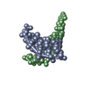
The crystal structure of Sanguinarine bound to DNA d(CGTACG)
PDB-5icf:
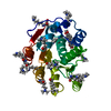
Crystal structure of (S)-norcoclaurine 6-O-methyltransferase with S-adenosyl-L-homocysteine and sanguinarine
PDB-5zbz:
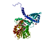
Crystal structure of the DEAD domain of Human eIF4A with sanguinarine
PDB-7c6q:
Novel natural PPARalpha agonist with a unique binding mode
