ジャーナル: J Biol Chem / 年: 2019 タイトル: The domain architecture of the protozoan protein J-DNA-binding protein 1 suggests synergy between base J DNA binding and thymidine hydroxylase activity. 著者: Athanassios Adamopoulos / Tatjana Heidebrecht / Jeroen Roosendaal / Wouter G Touw / Isabelle Q Phan / Jos Beijnen / Anastassis Perrakis / 要旨: J-DNA-binding protein 1 (JBP1) contributes to the biosynthesis and maintenance of base J (β-d-glucosyl-hydroxymethyluracil), an epigenetic modification of thymidine (T) confined to pathogenic ...J-DNA-binding protein 1 (JBP1) contributes to the biosynthesis and maintenance of base J (β-d-glucosyl-hydroxymethyluracil), an epigenetic modification of thymidine (T) confined to pathogenic protozoa such as and JBP1 has two known functional domains: an N-terminal T hydroxylase (TH) homologous to the 5-methylcytosine hydroxylase domain in TET proteins and a J-DNA-binding domain (JDBD) that resides in the middle of JBP1. Here, we show that removing JDBD from JBP1 results in a soluble protein (Δ-JDBD) with the N- and C-terminal regions tightly associated together in a well-ordered structure. We found that this Δ-JDBD domain retains TH activity but displays a 15-fold lower apparent rate of hydroxylation compared with JBP1. Small-angle X-ray scattering (SAXS) experiments on JBP1 and JDBD in the presence or absence of J-DNA and on Δ-JDBD enabled us to generate low-resolution three-dimensional models. We conclude that Δ-JDBD, and not the N-terminal region of JBP1 alone, is a distinct folding unit. Our SAXS-based model supports the notion that binding of JDBD specifically to J-DNA can facilitate T hydroxylation 12-14 bp downstream on the complementary strand of the J-recognition site. We postulate that insertion of the JDBD module into the Δ-JDBD scaffold during evolution provided a mechanism that synergized J recognition and T hydroxylation, ensuring inheritance of base J in specific sequence patterns following DNA replication in kinetoplastid parasites.
登録者
Nassos Adamopoulos (Netherlands Cancer Institute, Amsterdam, Netherlands)
 ムービー
ムービー コントローラー
 万見について


 データを開く
 基本情報
基本情報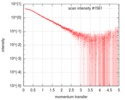
 試料
試料 引用
引用

 登録者
登録者 構造の表示
構造の表示 ダウンロードとリンク
 SASDGS2
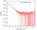
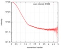
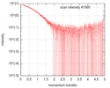
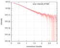
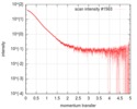
 / 線源: X-ray synchrotron / 波長: 0.1 Å / スペクトロメータ・検出器間距離: 2.872 mm


