 Movie
Movie Controller
[English] 日本語
 Yorodumi
- SASDFP8: Carbonic anhydrase 2 from bovine erythrocytes - SEC-SAXS coupled ... -
+ Open data

- Basic information
| Entry | Database: SASBDB / ID: SASDFP8 |
|---|---|
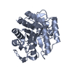
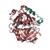
 Sample Sample | Carbonic anhydrase 2 from bovine erythrocytes - SEC-SAXS coupled to multiangle laser and quasi-elastic light scattering (MALLS and QELS)
|
| Function / homology |  Function and homology information Function and homology informationpositive regulation of dipeptide transmembrane transport / regulation of monoatomic anion transport / cyanamide hydratase / cyanamide hydratase activity / angiotensin-activated signaling pathway / regulation of intracellular pH / carbonic anhydrase / carbonate dehydratase activity / carbon dioxide transport / apical part of cell ...positive regulation of dipeptide transmembrane transport / regulation of monoatomic anion transport / cyanamide hydratase / cyanamide hydratase activity / angiotensin-activated signaling pathway / regulation of intracellular pH / carbonic anhydrase / carbonate dehydratase activity / carbon dioxide transport / apical part of cell / zinc ion binding / plasma membrane / cytoplasm Similarity search - Function |
| Biological species |  |
 Contact author Contact author |
|
- Structure visualization
| Structure viewer | Molecule: MolmilJmol/JSmol |
|---|
- Downloads & links
 SASDFP8


-Models
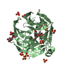
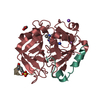
| Model #3213 |  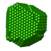 Type: dummy / Software: (SUPCOMB 23 (r9988)) / Radius of dummy atoms: 1.50 A / Symmetry: P1 Comment: Refined DAMMIN model obtained from the spatial alignment of 10 individual models (DAMSTART) Chi-square value: 1.020 / P-value: 0.448891  Search similar-shape structures of this assembly by Omokage search (details) Search similar-shape structures of this assembly by Omokage search (details) |
|---|---|
| Model #3214 |  Type: dummy / Software: (DAMFILT 5.0 (r10552)) / Radius of dummy atoms: 1.50 A / Symmetry: P1 Comment: DAMFILT spatially aligned and volume occupancy corrected (averaged) model from 10 individual models Chi-square value: 1.020 / P-value: 0.448891 Search similar-shape structures of this assembly by Omokage search (details) |
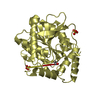
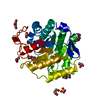
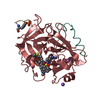
| Model #3215 |   Type: atomic / Symmetry: P1 Comment: Model-fit calculated using 30 harmonics and 300 points Chi-square value: 1.095 / P-value: 0.269894 Search similar-shape structures of this assembly by Omokage search (details) |
-Sample
| Sample | Name: Carbonic anhydrase 2 from bovine erythrocytes - SEC-SAXS coupled to multiangle laser and quasi-elastic light scattering (MALLS and QELS) Specimen concentration: 11.9 mg/ml |
|---|---|
| Buffer | Name: 50 mM HEPES, 150 mM NaCl, 2% v/v glycerol, / pH: 7 / Comment: Running buffer for SEC-SAXS |
| Entity #1768 | Name: CA2 / Type: protein / Description: Carbonic anhydrase 2 / Formula weight: 29.113 / Num. of mol.: 1 / Source: Bos taurus / References: UniProt: P00921 Sequence: MSHHWGYGKH NGPEHWHKDF PIANGERQSP VDIDTKAVVQ DPALKPLALV YGEATSRRMV NNGHSFNVEY DDSQDKAVLK DGPLTGTYRL VQFHFHWGSS DDQGSEHTVD RKKYAAELHL VHWNTKYGDF GTAAQQPDGL AVVGVFLKVG DANPALQKVL DALDSIKTKG ...Sequence: MSHHWGYGKH NGPEHWHKDF PIANGERQSP VDIDTKAVVQ DPALKPLALV YGEATSRRMV NNGHSFNVEY DDSQDKAVLK DGPLTGTYRL VQFHFHWGSS DDQGSEHTVD RKKYAAELHL VHWNTKYGDF GTAAQQPDGL AVVGVFLKVG DANPALQKVL DALDSIKTKG KSTDFPNFDP GSLLPNVLDY WTYPGSLTTP PLLESVTWIV LKEPISVSSQ QMLKFRTLNF NAEGEPELLM LANWRPAQPL KNRQVRGFPK |
-Experimental information
| Beam | Instrument name: PETRA III EMBL P12 / City: Hamburg / 国: Germany  / Type of source: X-ray synchrotron / Wavelength: 0.123982 Å / Dist. spec. to detc.: 3 mm / Type of source: X-ray synchrotron / Wavelength: 0.123982 Å / Dist. spec. to detc.: 3 mm | |||||||||||||||||||||||||||||||||
|---|---|---|---|---|---|---|---|---|---|---|---|---|---|---|---|---|---|---|---|---|---|---|---|---|---|---|---|---|---|---|---|---|---|---|
| Detector | Name: Pilatus 6M | |||||||||||||||||||||||||||||||||
| Scan |  Measurement date: Apr 5, 2019 / Storage temperature: 20 °C / Cell temperature: 20 °C / Exposure time: 1 sec. / Number of frames: 41 / Unit: 1/nm /
| |||||||||||||||||||||||||||||||||
| Distance distribution function P(R) | 
| |||||||||||||||||||||||||||||||||
| Result |  Comments: Carbonic anhydrase underwent pre-purification prior to SEC-SAXS using the following method. All procedures were performed at 4 oC. The protein (from Sigma; Gel Filtration Markers Kit ...Comments: Carbonic anhydrase underwent pre-purification prior to SEC-SAXS using the following method. All procedures were performed at 4 oC. The protein (from Sigma; Gel Filtration Markers Kit MWGF1000) was made to approximately 25 mg/ml in 25 mM HEPES, 50 mM NaCl, 5 mM urea, 1% v/v glycerol, pH 7. Approximately 200 μl of sample were loaded onto a Superdex 75 Increase 10/300 column (GE Healthcare) equilibrated in the same buffer (flow rate = 0.4 ml/min). Fractionated aliquots corresponding to the highest absorbing peak (estimated using UV A280 and UV A245 nm) were pooled and concentrated (3 kDa centrifuge spin filter) to a final concentration of 11.9 mg/ml (the concentration was determined from triplicate UV A280 measurements using an E0.1% of 1.732 (= 1 g/l) calculated from the amino acid sequence (ProtParam)). Approximately 50 μl aliquots were snap-frozen in liquid nitrogen then stored at -80oC prior to the SEC-SAXS analysis that was performed at room temperature in 50 mM HEPES, 150 mM NaCl, 2% v/v glycerol, pH 7. The Rg-correlation through the SEC-SAXS peak, the individual unsubtracted SEC-SAXS frames as well as the results from coupled MALLS and QELS analysis are included in the full entry zip archive. The quoted experimental molecular weight was determined using MALLS in combination with refractive-index (RI) measurements that were recorded from the same sample eluting from the column using a split-flow SEC-SAXS-light scattering configuration (Graewert et al., (2015) Sci. Reports. 5, 10734: doi: 10.1038/srep10734). The average hydrodynamic radius of the protein is 2.4 nm.
|
