 Movie
Movie Controller
[English] 日本語
 Yorodumi
- SASDEA6: Staphylococcus aureus N-acetylglucosamine-6-phosphate deacetylase... -
+ Open data

- Basic information
| Entry | Database: SASBDB / ID: SASDEA6 |
|---|---|
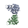
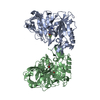
 Sample Sample | Staphylococcus aureus N-acetylglucosamine-6-phosphate deacetylase dimer
|
| Function / homology |  Function and homology information Function and homology informationN-acetylgalactosamine-6-phosphate deacetylase activity / N-acetylglucosamine-6-phosphate deacetylase / N-acetylglucosamine-6-phosphate deacetylase activity / N-acetylglucosamine metabolic process / metal ion binding Similarity search - Function |
| Biological species |  Staphylococcus aureus (strain USA300) (bacteria) Staphylococcus aureus (strain USA300) (bacteria) |
 Citation Citation | Journal: FEBS Lett / Year: 2019 Title: Functional and solution structure studies of amino sugar deacetylase and deaminase enzymes from Staphylococcus aureus. Authors: James S Davies / David Coombes / Christopher R Horne / F Grant Pearce / Rosmarie Friemann / Rachel A North / Renwick C J Dobson /    Abstract: N-Acetylglucosamine-6-phosphate deacetylase (NagA) and glucosamine-6-phosphate deaminase (NagB) are branch point enzymes that direct amino sugars into different pathways. For Staphylococcus aureus ...N-Acetylglucosamine-6-phosphate deacetylase (NagA) and glucosamine-6-phosphate deaminase (NagB) are branch point enzymes that direct amino sugars into different pathways. For Staphylococcus aureus NagA, analytical ultracentrifugation and small-angle X-ray scattering data demonstrate that it is an asymmetric dimer in solution. Initial rate experiments show hysteresis, which may be related to pathway regulation, and kinetic parameters similar to other bacterial isozymes. The enzyme binds two Zn ions and is not substrate inhibited, unlike the Escherichia coli isozyme. S. aureus NagB adopts a novel dimeric structure in solution and shows kinetic parameters comparable to other Gram-positive isozymes. In summary, these functional data and solution structures are of use for understanding amino sugar metabolism in S. aureus, and will inform the design of inhibitory molecules. |
 Contact author Contact author |
|
- Structure visualization
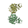
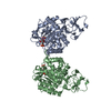
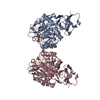
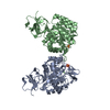
| Structure viewer | Molecule: MolmilJmol/JSmol |
|---|
- Downloads & links
-Data source
| SASBDB page |  SASDEA6 SASDEA6 |
|---|
-Related structure data
| Related structure data | C: citing same article ( |
|---|---|
| Similar structure data |


-External links
| Related items in Molecule of the Month |
|---|


-Models
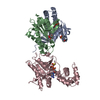
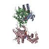
| Model #2491 |  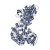 Type: atomic / Software: (2.0.6) Comment: Structure of NagA isozyme from B subtilis fit to data. Generated using symmetry mates. Chi-square value: 0.207  Search similar-shape structures of this assembly by Omokage search (details) Search similar-shape structures of this assembly by Omokage search (details) |
|---|
-Sample
| Sample | Name: Staphylococcus aureus N-acetylglucosamine-6-phosphate deacetylase dimer Specimen concentration: 6 mg/ml |
|---|---|
| Buffer | Name: 20 mM Tris-HCl 150 mM NaCl / pH: 8 |
| Entity #1330 | Name: NagA / Type: protein / Description: N-acetylglucosamine-6-phosphate deacetylase / Formula weight: 43.115 / Num. of mol.: 2 / Source: Staphylococcus aureus (strain USA300) / References: UniProt: A0A0H2XGG9 Sequence: MSELIIYNGK VYTEDGKIDN GYIHVKDGQI VAIGEVDDKA AIDNDTTNKI QVIDAKGHHV LPGFIDIHIH GGYGQDAMDG SYDGLKYLSE NLLSEGTTSY LATTMTQSTD KIDNALTNIA KYEAEQDVHN AAEIVGIHLE GPFISENKVG AQHPQYVVRP FIDKIKHFQE ...Sequence: MSELIIYNGK VYTEDGKIDN GYIHVKDGQI VAIGEVDDKA AIDNDTTNKI QVIDAKGHHV LPGFIDIHIH GGYGQDAMDG SYDGLKYLSE NLLSEGTTSY LATTMTQSTD KIDNALTNIA KYEAEQDVHN AAEIVGIHLE GPFISENKVG AQHPQYVVRP FIDKIKHFQE TANGLIKIMT FAPEVEGAKE ALETYKDDII FSIGHTVATY EEAVEAVERG AKHVTHLYNA ATPFQHREPG VFGAAWLNDA LHTEMIVDGT HSHPASVAIA YRMKGNERFY LITDAMRAKG MPEGEYDLGG QKVTVQSQQA RLANGALAGS ILKMNHGLRN LISFTGDTLD HLWRVTSLNQ AIALGIDDRK GSIKVNKDAD LVILDDDMNV KSTIKQGKVH TFS |
-Experimental information
| Beam | Instrument name: Australian Synchrotron SAXS/WAXS / City: Melbourne / 国: Australia / Shape: Point / Type of source: X-ray synchrotron / Wavelength: 0.10332 Å / Dist. spec. to detc.: 1.6 mm | ||||||||||||||||||||||||||||||
|---|---|---|---|---|---|---|---|---|---|---|---|---|---|---|---|---|---|---|---|---|---|---|---|---|---|---|---|---|---|---|---|
| Detector | Name: Pilatus 1M / Type: Dectris / Pixsize x: 172 mm | ||||||||||||||||||||||||||||||
| Scan |  Measurement date: Apr 26, 2016 / Storage temperature: 4 °C / Cell temperature: 20 °C / Exposure time: 1 sec. / Number of frames: 999 / Unit: 1/A /
| ||||||||||||||||||||||||||||||
| Distance distribution function P(R) | 
| ||||||||||||||||||||||||||||||
| Result | 
|
