 ムービー
ムービー コントローラー

+ データを開く

- 基本情報
基本情報
| 登録情報 | データベース: SASBDB / ID: SASDD83 |
|---|---|
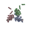
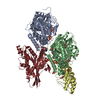
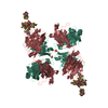
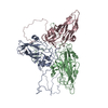
 試料 試料 | Apolipoprotein D (ApoD) tetramer
|
| 機能・相同性 |  機能・相同性情報 機能・相同性情報negative regulation of lipoprotein lipid oxidation / Transport of fatty acids / negative regulation of T cell migration / negative regulation of smooth muscle cell-matrix adhesion / peripheral nervous system axon regeneration / tissue regeneration / negative regulation of focal adhesion assembly / lipid transporter activity / negative regulation of monocyte chemotactic protein-1 production / negative regulation of platelet-derived growth factor receptor signaling pathway ...negative regulation of lipoprotein lipid oxidation / Transport of fatty acids / negative regulation of T cell migration / negative regulation of smooth muscle cell-matrix adhesion / peripheral nervous system axon regeneration / tissue regeneration / negative regulation of focal adhesion assembly / lipid transporter activity / negative regulation of monocyte chemotactic protein-1 production / negative regulation of platelet-derived growth factor receptor signaling pathway / cholesterol binding / negative regulation of protein import into nucleus / response to axon injury / NR1H3 & NR1H2 regulate gene expression linked to cholesterol transport and efflux / cytosolic ribosome / negative regulation of cytokine production involved in inflammatory response / response to reactive oxygen species / negative regulation of smooth muscle cell proliferation / brain development / lipid metabolic process / glucose metabolic process / angiogenesis / neuronal cell body / dendrite / perinuclear region of cytoplasm / endoplasmic reticulum / extracellular space / extracellular exosome / extracellular region / cytoplasm 類似検索 - 分子機能 |
| 生物種 |  Homo sapiens (ヒト) Homo sapiens (ヒト) |
 引用 引用 | ジャーナル: J Struct Biol / 年: 2018 タイトル: Identification of a novel tetrameric structure for human apolipoprotein-D. 著者: Claudia S Kielkopf / Jason K K Low / Yee-Foong Mok / Surabhi Bhatia / Tony Palasovski / Aaron J Oakley / Andrew E Whitten / Brett Garner / Simon H J Brown /  要旨: Apolipoprotein-D is a 25 kDa glycosylated member of the lipocalin family that folds into an eight-stranded β-barrel with a single adjacent α-helix. Apolipoprotein-D specifically binds a range of ...Apolipoprotein-D is a 25 kDa glycosylated member of the lipocalin family that folds into an eight-stranded β-barrel with a single adjacent α-helix. Apolipoprotein-D specifically binds a range of small hydrophobic ligands such as progesterone and arachidonic acid and has an antioxidant function that is in part due to the reduction of peroxidised lipids by methionine-93. Therefore, apolipoprotein-D plays multiple roles throughout the body and is protective in Alzheimer's disease, where apolipoprotein-D overexpression reduces the amyloid-β burden in Alzheimer's disease mouse models. Oligomerisation is a common feature of lipocalins that can influence ligand binding. The native structure of apolipoprotein-D, however, has not been conclusively defined. Apolipoprotein-D is generally described as a monomeric protein, although it dimerises when reducing peroxidised lipids. Here, we investigated the native structure of apolipoprotein-D derived from plasma, breast cyst fluid (BCF) and cerebrospinal fluid. In plasma and cerebrospinal fluid, apolipoprotein-D was present in high-molecular weight complexes, potentially in association with lipoproteins. In contrast, apolipoprotein-D in BCF formed distinct oligomeric species. We assessed apolipoprotein-D oligomerisation using native apolipoprotein-D purified from BCF and a suite of complementary methods, including multi-angle laser light scattering, analytical ultracentrifugation and small-angle X-ray scattering. Our analyses showed that apolipoprotein-D predominantly forms a ∼95 to ∼100 kDa tetramer. Small-angle X-ray scattering analysis confirmed these findings and provided a structural model for apolipoprotein-D tetramer. These data indicate apolipoprotein-D rarely exists as a free monomer under physiological conditions and provide insights into novel native structures of apolipoprotein-D and into oligomerisation behaviour in the lipocalin family. |
 登録者 登録者 |
|
- 構造の表示
構造の表示
| 構造ビューア | 分子: MolmilJmol/JSmol |
|---|
- ダウンロードとリンク
 SASDD83
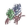


-モデル
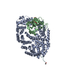
| モデル #2054 |   タイプ: atomic / ダミー原子の半径: 1.90 A / 対称性: P222 コメント: The restraints used are 32 Ang between: a)K55-K55; b) K156-K156; c) K155-K156 and; d) K144-K156 カイ2乗値: 1.28  Omokage検索でこの集合体の類似形状データを探す (詳細) Omokage検索でこの集合体の類似形状データを探す (詳細) |
|---|---|
| モデル #2055 |  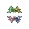 タイプ: atomic / ダミー原子の半径: 1.90 A / 対称性: P222 コメント: The restraints used are 32 Ang between: a) K55-K55; b) K156-K156; c) K155-K156 and; d) K144-K156. カイ2乗値: 1.26 Omokage検索でこの集合体の類似形状データを探す (詳細) |
-試料
| 試料 | 名称: Apolipoprotein D (ApoD) tetramer |
|---|---|
| バッファ | 名称: 50 mM Na Phosphate, 150 mM NaCl, 3% glycerol / pH: 7.4 |
| 要素 #956 | 名称: ApoD / タイプ: protein / 記述: Apolipoprotein D / 分子量: 19.303 / 分子数: 4 / 由来: Homo sapiens / 参照: UniProt: P05090 配列: QAFHLGKCPN PPVQENFDVN KYLGRWYEIE KIPTTFENGR CIQANYSLME NGKIKVLNQE LRADGTVNQI EGEATPVNLT EPAKLEVKFS WFMPSAPYWI LATDYENYAL VYSCTCIIQL FHVDFAWILA RNPNLPPETV DSLKNILTSN NIDVKKMTVT DQVNCPKLS |
-実験情報
| ビーム | 設備名称: Australian Synchrotron SAXS/WAXS / 地域: Melbourne / 国: Australia / 形状: Point / 線源: X-ray synchrotron / 波長: 0.10322 Å | |||||||||||||||||||||||||||||||||||||||
|---|---|---|---|---|---|---|---|---|---|---|---|---|---|---|---|---|---|---|---|---|---|---|---|---|---|---|---|---|---|---|---|---|---|---|---|---|---|---|---|---|
| 検出器 | 名称: Pilatus 1M / Pixsize x: 172 mm | |||||||||||||||||||||||||||||||||||||||
| スキャン | 
| |||||||||||||||||||||||||||||||||||||||
| 距離分布関数 P(R) | 
| |||||||||||||||||||||||||||||||||||||||
| 結果 |  コメント: Additional SEC-SAXS information: SEC-column: GE Healthcare Superdex S200 5/150; Loading concentration (measured using BCA assay): 7.5 mg/ml; Injection volume : 50 µl; Flow rate: 0.45 ...コメント: Additional SEC-SAXS information: SEC-column: GE Healthcare Superdex S200 5/150; Loading concentration (measured using BCA assay): 7.5 mg/ml; Injection volume : 50 µl; Flow rate: 0.45 ml/min; Number of frames averaged over the elution peak: 15. The starting pdb model was the apo-form of apoD with modelled glycans (Oakley et al. 2012). The refined SASREF CV model incorporated contact restraints derived from crosslinking mass spectrometry (crosslinker BS3) that are included in the full entry zip archive (32 Ang between: a, K55-K55; b, K156-K156; c, K155-K156 and; d, K144-K156.)
|
