 Movie
Movie Controller

+ Open data

- Basic information
| Entry | Database: SASBDB / ID: SASDBY2 |
|---|---|
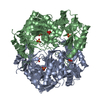
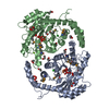
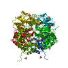
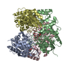
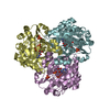
 Sample Sample | Inorganic pyrophosphatase (PPase) from E. coli
|
| Function / homology |  Function and homology information Function and homology informationinorganic triphosphate phosphatase activity / inorganic diphosphatase / inorganic diphosphate phosphatase activity / phosphate-containing compound metabolic process / magnesium ion binding / zinc ion binding / membrane / cytosol Similarity search - Function |
| Biological species |  |
 Citation Citation | Journal: PLoS One / Year: 2016 Title: X-Ray Solution Scattering Study of Four Escherichia coli Enzymes Involved in Stationary-Phase Metabolism. Authors: Liubov A Dadinova / Eleonora V Shtykova / Petr V Konarev / Elena V Rodina / Natalia E Snalina / Natalia N Vorobyeva / Svetlana A Kurilova / Tatyana I Nazarova / Cy M Jeffries / Dmitri I Svergun /   Abstract: The structural analyses of four metabolic enzymes that maintain and regulate the stationary growth phase of Escherichia coli have been performed primarily drawing on the results obtained from ...The structural analyses of four metabolic enzymes that maintain and regulate the stationary growth phase of Escherichia coli have been performed primarily drawing on the results obtained from solution small angle X-ray scattering (SAXS) and other structural techniques. The proteins are (i) class I fructose-1,6-bisphosphate aldolase (FbaB); (ii) inorganic pyrophosphatase (PPase); (iii) 5-keto-4-deoxyuronate isomerase (KduI); and (iv) glutamate decarboxylase (GadA). The enzyme FbaB, that until now had an unknown structure, is predicted to fold into a TIM-barrel motif that form globular protomers which SAXS experiments show associate into decameric assemblies. In agreement with previously reported crystal structures, PPase forms hexamers in solution that are similar to the previously reported X-ray crystal structure. Both KduI and GadA that are responsible for carbohydrate (pectin) metabolism and acid stress responses, respectively, form polydisperse mixtures consisting of different oligomeric states. Overall the SAXS experiments yield additional insights into shape and organization of these metabolic enzymes and further demonstrate the utility of hybrid methods, i.e., solution SAXS combined with X-ray crystallography, bioinformatics and predictive 3D-structural modeling, as tools to enrich structural studies. The results highlight the structural complexity that the protein components of metabolic networks may adopt which cannot be fully captured using individual structural biology techniques. |
 Contact author Contact author |
|
- Structure visualization
| Structure viewer | Molecule: MolmilJmol/JSmol |
|---|
- Downloads & links
 SASDBY2
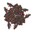


-Models
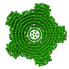
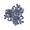
| Model #418 |   Type: dummy / Radius of dummy atoms: 2.00 A / Symmetry: P32 / Chi-square value: 1.032  Search similar-shape structures of this assembly by Omokage search (details) Search similar-shape structures of this assembly by Omokage search (details) |
|---|---|
| Model #419 |   Type: atomic / Radius of dummy atoms: 1.90 A / Symmetry: P32 / Chi-square value: 1.437 Search similar-shape structures of this assembly by Omokage search (details) |
| Model #420 |  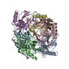 Type: atomic / Chi-square value: 5.180 Search similar-shape structures of this assembly by Omokage search (details) |
-Sample
| Sample | Name: Inorganic pyrophosphatase (PPase) from E. coli / Specimen concentration: 1.40-10.80 |
|---|---|
| Buffer | Name: 50 mM Tris 10 mM NaCl / Concentration: 50.00 mM / pH: 7.5 / Composition: 10 mM NaCl |
| Entity #279 | Name: PPase / Type: protein / Description: Inorganic pyrophosphatase (PPase) from E. coli / Formula weight: 19.56 / Num. of mol.: 6 / Source: Escherichia coli / References: UniProt: P0A7A9 Sequence: SLLNVPAGKD LPEDIYVVIE IPANADPIKY EIDKESGALF VDRFMSTAMF YPCNYGYINH TLSLDGDPVD VLVPTPYPLQ PGSVTRCRPV GVLKMTDEAG EDAKLVAVPH SKLSKEYDHI KDVNDLPELL KAQIAHFFEH YKDLEKGKWV KVEGWENAEA AKAEIVASFE RAKNK |
-Experimental information
| Beam | Instrument name: PETRA III EMBL P12 / City: Hamburg / 国: Germany / Type of source: X-ray synchrotron / Wavelength: 0.12 Å / Dist. spec. to detc.: 3.1 mm | ||||||||||||||||||||||||||||||||||||
|---|---|---|---|---|---|---|---|---|---|---|---|---|---|---|---|---|---|---|---|---|---|---|---|---|---|---|---|---|---|---|---|---|---|---|---|---|---|
| Detector | Name: Pilatus 2M | ||||||||||||||||||||||||||||||||||||
| Scan | 
| ||||||||||||||||||||||||||||||||||||
| Distance distribution function P(R) | 
| ||||||||||||||||||||||||||||||||||||
| Result |  Comments: The models displayed above are, from top to bottom: 1) An individual DAMMIN dummy atom model; 2) SASREF rigid body model with the associated CRYSOL30 fit to the data and; 3) The PPase ...Comments: The models displayed above are, from top to bottom: 1) An individual DAMMIN dummy atom model; 2) SASREF rigid body model with the associated CRYSOL30 fit to the data and; 3) The PPase crystal structure fit to the data (derived from Protein data bank entry 2AUU). All of the individual DAMMIN models, associated fits and averaging can be found in the zip archive for this entry.
|
