 Movie
Movie Controller
[English] 日本語
 Yorodumi
- SASDBU4: Vaccinia primase D5 protein fragment containing the D5N and helic... -
+ Open data

- Basic information
| Entry | Database: SASBDB / ID: SASDBU4 |
|---|---|
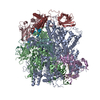
 Sample Sample | Vaccinia primase D5 protein fragment containing the D5N and helicase domain
|
| Function / homology |  Function and homology information Function and homology informationhelicase activity / Hydrolases; Acting on acid anhydrides; Acting on acid anhydrides to facilitate cellular and subcellular movement / host cell cytoplasm / hydrolase activity / ATP binding Similarity search - Function |
| Biological species |  Vaccinia virus Vaccinia virus |
 Citation Citation | Journal: J Virol / Year: 2016 Title: Domain Organization of Vaccinia Virus Helicase-Primase D5. Authors: Stephanie Hutin / Wai Li Ling / Adam Round / Gregory Effantin / Stefan Reich / Frédéric Iseni / Nicolas Tarbouriech / Guy Schoehn / Wim Pascal Burmeister /   Abstract: Poxviridae are viruses with a large linear double-stranded DNA genome coding for up to 250 open reading frames and a fully cytoplasmic replication. The double-stranded DNA genome is covalently ...Poxviridae are viruses with a large linear double-stranded DNA genome coding for up to 250 open reading frames and a fully cytoplasmic replication. The double-stranded DNA genome is covalently circularized at both ends. Similar structures of covalently linked extremities of the linear DNA genome are found in the African swine fever virus (asfarvirus) and in the Phycodnaviridae We are studying the machinery which replicates this peculiar genome structure. From our work with vaccinia virus, we give first insights into the overall structure and function of the essential poxvirus virus helicase-primase D5 and show that the active helicase domain of D5 builds a hexameric ring structure. This hexamer has ATPase and, more generally, nucleoside triphosphatase activities that are indistinguishable from the activities of full-length D5 and that are independent of the nature of the base. In addition, hexameric helicase domains bind tightly to single- and double-stranded DNA. Still, the monomeric D5 helicase construct truncated within the D5N domain leads to a well-defined structure, but it does not have ATPase or DNA-binding activity. This shows that the full D5N domain has to be present for hexamerization. This allowed us to assign a function to the D5N domain which is present not only in D5 but also in other viruses of the nucleocytoplasmic large DNA virus (NCLDV) clade. The primase domain and the helicase domain were structurally analyzed via a combination of small-angle X-ray scattering and, when appropriate, electron microscopy, leading to consistent low-resolution models of the different proteins. IMPORTANCE: Since the beginning of the 1980s, research on the vaccinia virus replication mechanism has basically stalled due to the absence of structural information. As a result, this important ...IMPORTANCE: Since the beginning of the 1980s, research on the vaccinia virus replication mechanism has basically stalled due to the absence of structural information. As a result, this important class of pathogens is less well understood than most other viruses. This lack of information concerns in general viruses of the NCLDV clade, which use a superfamily 3 helicase for replication, as do poxviruses. Here we provide for the first time information about the domain structure and DNA-binding activity of D5, the poxvirus helicase-primase. This result not only refines the current model of the poxvirus replication fork but also will lead in the long run to a structural basis for antiviral drug design. |
 Contact author Contact author |
|
- Structure visualization
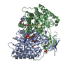
| Structure viewer | Molecule: MolmilJmol/JSmol |
|---|
- Downloads & links
-Data source
| SASBDB page |  SASDBU4 SASDBU4 |
|---|
-Related structure data
| Similar structure data |
|---|


-External links
| Related items in Molecule of the Month |
|---|


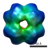
-Models
| Model #534 |  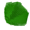 Type: dummy / Software: DAMMIF (r3709) / Radius of dummy atoms: 3.00 A / Chi-square value: 1.096209 / P-value: 0.000004  Search similar-shape structures of this assembly by Omokage search (details) Search similar-shape structures of this assembly by Omokage search (details) |
|---|
-Sample
| Sample | Name: Vaccinia primase D5 protein fragment containing the D5N and helicase domain Specimen concentration: 1.42-2.20 |
|---|---|
| Buffer | Name: Tris / Concentration: 20.00 mM / pH: 7 / Composition: 150 mM NaCL, 5% glycerol, 1mM DTT |
| Entity #342 | Name: Vaccinia D5 323-785 / Type: protein Description: Primase D5 protein fragment containing the D5N and helicase domain Formula weight: 53.48 / Num. of mol.: 6 / Source: Vaccinia virus / References: UniProt: P21010 Sequence: GAMGNKLFNI AQRILDTNSV LLTERGDYIV WINNSWKFNS EEPLITKLIL SIRHQLPKEY SSELLCPRKR KTVEANIRDM LVDSVETDTY PDKLPFKNGV LDLVDGMFYS GDDAKKYTCT VSTGFKFDDT KFVEDSPEME ELMNIINDIQ PLTDENKKNR ELYEKTLSSC ...Sequence: GAMGNKLFNI AQRILDTNSV LLTERGDYIV WINNSWKFNS EEPLITKLIL SIRHQLPKEY SSELLCPRKR KTVEANIRDM LVDSVETDTY PDKLPFKNGV LDLVDGMFYS GDDAKKYTCT VSTGFKFDDT KFVEDSPEME ELMNIINDIQ PLTDENKKNR ELYEKTLSSC LCGATKGCLT FFFGETATGK STTKRLLKSA IGDLFVETGQ TILTDVLDKG PNPFIANMHL KRSVFCSELP DFACSGSKKI RSDNIKKLTE PCVIGRPCFS NKINNRNHAT IIIDTNYKPV FDRIDNALMR RIAVVRFRTH FSQPSGREAA ENNDAYDKVK LLDEGLDGKI QNNRYRFAFL YLLVKWYKKY HVPIMKLYPT PEEIPDFAFY LKIGTLLVSS SVKHIPLMTD LSKKGYILYD NVVTLPLTTF QQKISKYFNS RLFGHDIESF INRHKKFANV SDEYLQYIFI EDISSP |
-Experimental information
| Beam | Instrument name: ESRF BM29 / City: Grenoble / 国: France / Type of source: X-ray synchrotron / Wavelength: 0.93 Å / Dist. spec. to detc.: 2.43 mm | |||||||||||||||||||||||||||||||||
|---|---|---|---|---|---|---|---|---|---|---|---|---|---|---|---|---|---|---|---|---|---|---|---|---|---|---|---|---|---|---|---|---|---|---|
| Detector | Name: Pilatus 1M | |||||||||||||||||||||||||||||||||
| Scan | 
| |||||||||||||||||||||||||||||||||
| Distance distribution function P(R) | 
| |||||||||||||||||||||||||||||||||
| Result | 
|
