 Movie
Movie Controller
 Structure viewers
 About Yorodumi Papers


+Search query
-Structure paper
| Title | Domain Organization of Vaccinia Virus Helicase-Primase D5. |
|---|---|
| Journal, issue, pages | J Virol, Vol. 90, Issue 9, Page 4604-4613, Year 2016 |
| Publish date | Apr 14, 2016 |
 Authors Authors | Stephanie Hutin / Wai Li Ling / Adam Round / Gregory Effantin / Stefan Reich / Frédéric Iseni / Nicolas Tarbouriech / Guy Schoehn / Wim Pascal Burmeister /   |
| PubMed Abstract | Poxviridae are viruses with a large linear double-stranded DNA genome coding for up to 250 open reading frames and a fully cytoplasmic replication. The double-stranded DNA genome is covalently ...Poxviridae are viruses with a large linear double-stranded DNA genome coding for up to 250 open reading frames and a fully cytoplasmic replication. The double-stranded DNA genome is covalently circularized at both ends. Similar structures of covalently linked extremities of the linear DNA genome are found in the African swine fever virus (asfarvirus) and in the Phycodnaviridae We are studying the machinery which replicates this peculiar genome structure. From our work with vaccinia virus, we give first insights into the overall structure and function of the essential poxvirus virus helicase-primase D5 and show that the active helicase domain of D5 builds a hexameric ring structure. This hexamer has ATPase and, more generally, nucleoside triphosphatase activities that are indistinguishable from the activities of full-length D5 and that are independent of the nature of the base. In addition, hexameric helicase domains bind tightly to single- and double-stranded DNA. Still, the monomeric D5 helicase construct truncated within the D5N domain leads to a well-defined structure, but it does not have ATPase or DNA-binding activity. This shows that the full D5N domain has to be present for hexamerization. This allowed us to assign a function to the D5N domain which is present not only in D5 but also in other viruses of the nucleocytoplasmic large DNA virus (NCLDV) clade. The primase domain and the helicase domain were structurally analyzed via a combination of small-angle X-ray scattering and, when appropriate, electron microscopy, leading to consistent low-resolution models of the different proteins. IMPORTANCE: Since the beginning of the 1980s, research on the vaccinia virus replication mechanism has basically stalled due to the absence of structural information. As a result, this important class of pathogens is less well understood than most other viruses. This lack of information concerns in general viruses of the NCLDV clade, which use a superfamily 3 helicase for replication, as do poxviruses. Here we provide for the first time information about the domain structure and DNA-binding activity of D5, the poxvirus helicase-primase. This result not only refines the current model of the poxvirus replication fork but also will lead in the long run to a structural basis for antiviral drug design. |
 External links External links | J Virol / PubMed:26912611 / PubMed Central |
| Methods | SAS (X-ray synchrotron) |
| Structure data |  SASDBU4: |
| Source |
|

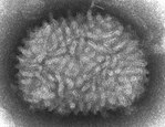
 Vaccinia virus