 Movie
Movie Controller

+ Open data

- Basic information
| Entry | Database: SASBDB / ID: SASDBN2 |
|---|---|
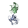
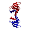
 Sample Sample | Human linear diubiquitin
|
| Function / homology |  Function and homology information Function and homology informationhypothalamus gonadotrophin-releasing hormone neuron development / female meiosis I / fat pad development / seminiferous tubule development / female gonad development / male meiosis I / energy homeostasis / mitochondrial outer membrane / nucleus Similarity search - Function |
| Biological species |  Homo sapiens (human) Homo sapiens (human) |
 Citation Citation | Journal: Acta Crystallogr D Struct Biol / Year: 2016 Title: New conformations of linear polyubiquitin chains from crystallographic and solution-scattering studies expand the conformational space of polyubiquitin. Authors: Trung Thanh Thach / Donghyuk Shin / Seungsu Han / Sangho Lee /  Abstract: The conformational flexibility of linkage-specific polyubiquitin chains enables ubiquitylated proteins and their receptors to be involved in a variety of cellular processes. Linear or Met1-linked ...The conformational flexibility of linkage-specific polyubiquitin chains enables ubiquitylated proteins and their receptors to be involved in a variety of cellular processes. Linear or Met1-linked polyubiquitin chains, associated with nondegradational cellular signalling pathways, have been known to adopt multiple conformations from compact to extended conformations. However, the extent of such conformational flexibility remains open. Here, the crystal structure of linear Ub2 was determined in a more compact conformation than that of the previously known structure (PDB entry 3axc). The two structures differ significantly from each other, as shown by an r.m.s.d. between C(α) atoms of 3.1 Å. The compactness of the linear Ub2 structure in comparison with PDB entry 3axc is supported by smaller values of the radius of gyration (Rg; 18 versus 18.9 Å) and the maximum interatomic distance (Dmax; 55.5 versus 57.8 Å). Extra intramolecular hydrogen bonds formed among polar residues between the distal and proximal ubiquitin moieties seem to contribute to stabilization of the compact conformation of linear Ub2. An ensemble of three semi-extended and extended conformations of linear Ub2 was also observed by small-angle X-ray scattering (SAXS) analysis in solution. In addition, the conformational heterogeneity in linear polyubiquitin chains is clearly manifested by SAXS analyses of linear Ub3 and Ub4: at least three distinct solution conformations are observed in each chain, with the linear Ub3 conformations being compact. The results expand the extent of conformational space of linear polyubiquitin chains and suggest that changes in the conformational ensemble may be pivotal in mediating multiple signalling pathways. |
 Contact author Contact author |
|
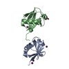
- Structure visualization
| Structure viewer | Molecule: MolmilJmol/JSmol |
|---|
- Downloads & links
 SASDBN2
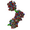


-Models
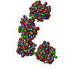
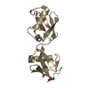
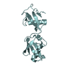
| Model #405 |   Type: mix / Software: EOM / Radius of dummy atoms: 1.90 A / Chi-square value: 0.278343821889  Search similar-shape structures of this assembly by Omokage search (details) Search similar-shape structures of this assembly by Omokage search (details) |
|---|---|
| Model #406 |  Type: mix / Software: EOM / Radius of dummy atoms: 1.90 A / Chi-square value: 0.278343821889 Search similar-shape structures of this assembly by Omokage search (details) |
-Sample
| Sample | Name: Human linear diubiquitin / Specimen concentration: 1.15-4.60 |
|---|---|
| Buffer | Name: 50 mM Tris / pH: 7.5 / Composition: 150 mM NaCl, 1 mM MgCl2 |
| Entity #268 | Type: protein / Description: Linear di-ubiquitin / Formula weight: 17.111 / Num. of mol.: 1 / Source: Homo sapiens / References: UniProt: Q5U5U6 Sequence: MQIFVKTLTG KTITLEVEPS DTIENVKAKI QDKEGIPPDQ QRLIFAGKQL EDGRTLSDYN IQKESTLHLV LRLRGGMQIF VKTLTGKTIT LEVEPSDTIE NVKAKIQDKE GIPPDQQRLI FAGKQLEDGR TLSDYNIQKE STLHLVLRLR GG |
-Experimental information
| Beam | Instrument name: Pohang Accelerator Laboratory 5C / City: Pohang / 国: South Korea / Type of source: X-ray synchrotron / Wavelength: 1.907 Å / Dist. spec. to detc.: 1.4 mm | |||||||||||||||||||||||||||||||||
|---|---|---|---|---|---|---|---|---|---|---|---|---|---|---|---|---|---|---|---|---|---|---|---|---|---|---|---|---|---|---|---|---|---|---|
| Detector | Name: ADSC Quantum 315 / Type: CCD | |||||||||||||||||||||||||||||||||
| Scan | 
| |||||||||||||||||||||||||||||||||
| Distance distribution function P(R) | 
| |||||||||||||||||||||||||||||||||
| Result |  Comments: Note: The displayed results show a merged data set derived from a concentration series. Data were normalised to protein concentration and the high angle data from highest concentration and ...Comments: Note: The displayed results show a merged data set derived from a concentration series. Data were normalised to protein concentration and the high angle data from highest concentration and low angle data from lowest concentration were merged to generate final SAXS profile. Because of the flexibility of linear-diubiquitin molecule, ensemble optimisation method, EOM, was performed. The models shown above are two diubiquitin examples from the final EOM output. All EOM models, the radius of gyration distribution and maximum particle dimension distribution can be found in the full entry zip archive.
|
