 Movie
Movie Controller

 Yorodumi
+ Open data

- Basic information
| Entry | 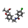 Database: PDB chemical components / ID: PEM Database: PDB chemical components / ID: PEM |
|---|---|
| Name | Name: |
-Chemical information
| Composition |  | ||||
|---|---|---|---|---|---|
| Others | Type: NON-POLYMER / PDB classification: HETAIN / Three letter code: PEM / Ideal coordinates details: OpenEye/OEToolkits V1.4.2 / Model coordinates PDB-ID: 1IWH | ||||
| History |
| ||||
 External links External links | UniChem / ChemSpider / BindingDB / Brenda / ChEBI / ChEMBL / CompTox / DrugBank / GtoPharmacology / HMDB / PubChem / SureChEMBL / Wikipedia search / Google search |
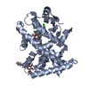
- Structure visualization
| Structure viewer | Molecule:  MolmilJmol/JSmol MolmilJmol/JSmol |
|---|
-Details
-SMILES
| ACDLabs 10.04 | | CACTVS 3.341 | OpenEye OEToolkits 1.5.0 | |
|---|
-SMILES CANONICAL
| CACTVS 3.341 | | OpenEye OEToolkits 1.5.0 | |
|---|
-InChI
| InChI 1.03 |
|---|
-InChIKey
| InChI 1.03 |
|---|
-SYSTEMATIC NAME
| ACDLabs 10.04 | | OpenEye OEToolkits 1.5.0 | |
|---|
-PDB entries
Showing all 6 items
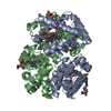
PDB-1iwh: 
Crystal Structure of Horse Carbonmonoxyhemoglobin-Bezafibrate Complex at 1.55A Resolution: A Novel Allosteric Binding Site in R-State Hemoglobin
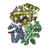
PDB-5x2s:
Direct Observation of Conformational Population Shifts in Hemoglobin: Crystal Structure of Half-Liganded Hemoglobin after Adding 4 mM bezafibrate pH 6.5.
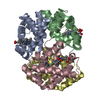
PDB-5x2t:
Direct Observation of Conformational Population Shifts in Hemoglobin: Crystal Structure of Half-Liganded Hemoglobin after Adding 4 mM bezafibrate pH 7.2.

PDB-7bpz:
X-ray structure of human PPARalpha ligand binding domain-bezafibrate-SRC1 coactivator peptide co-crystals obtained by soaking
PDB-7wgl:
X-ray structure of human PPAR delta ligand binding domain-bezafibrate co-crystals obtained by co-crystallization

PDB-7wgo:
X-ray structure of human PPAR gamma ligand binding domain-bezafibrate co-rystals obtained by co-crystallization
