 Movie
Movie Controller


+ Open data

- Basic information
| Entry | Database: PDB / ID: 9xqm | |||||||||
|---|---|---|---|---|---|---|---|---|---|---|
| Title | The structure of PldB-PA5088 complex state_2 | |||||||||
 Components Components |
| |||||||||
 Keywords Keywords | IMMUNE SYSTEM / Inhibitor / Complex | |||||||||
| Function / homology |  Function and homology information Function and homology information | |||||||||
| Biological species |  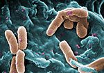 Pseudomonas aeruginosa (bacteria) Pseudomonas aeruginosa (bacteria) | |||||||||
| Method | ELECTRON MICROSCOPY / single particle reconstruction / cryo EM / Resolution: 3.56 Å | |||||||||
 Authors Authors | Yang, J.W. / Yang, X.Y. / Li, Z.Q. | |||||||||
| Funding support |  China, 1items China, 1items
| |||||||||
 Citation Citation | Journal: Commun Biol / Year: 2026 Title: Structural basis of cooperative neutralization of the PldB toxin by immunity proteins in Pseudomonas aeruginosa. Authors: Xiaoyun Yang / Jiawen Yang / Hong Wang / Xiuhua Liu / Zongqiang Li / Abstract: PldB, a type VI secretion system-dependent phospholipase D (PLD) effector secreted by multidrug-resistant Pseudomonas aeruginosa, alters host cell membrane permeability and facilitates pathogen ...PldB, a type VI secretion system-dependent phospholipase D (PLD) effector secreted by multidrug-resistant Pseudomonas aeruginosa, alters host cell membrane permeability and facilitates pathogen internalization. Its cytotoxic activity is neutralized by three cognate immunity proteins-PA5086, PA5087, and PA5088-which protect the bacterium from self-intoxication. However, the underlying mechanism remains unclear. Through quantitative and qualitative analyses, we demonstrate that these three immunity proteins function cooperatively to inhibit PldB toxicity. Cryogenic electron microscopy of the PldB-PA5088 complex reveals that PA5088 binds to the HKD2 domain of PldB primarily through electrostatic interactions, markedly reducing the volume of its active center. Interaction studies using domain‑specific truncated PldB variants, together with enzyme activity assays, identify distinct copy numbers and binding regions for PA5086, PA5087, and PA5088 in their association with PldB. Collectively, our findings provide mechanistic insights into immunity protein-mediated neutralization of PldB toxicity, offering a potential foundation for designing PLD-targeting therapeutics against P. aeruginosa infection. | |||||||||
| History |
|
- Structure visualization
| Structure viewer | Molecule: MolmilJmol/JSmol |
|---|
- Downloads & links
-Download
| PDBx/mmCIF format | 9xqm.cif.gz | 143.7 KB | Display | PDBx/mmCIF format |
|---|---|---|---|---|
| PDB format | pdb9xqm.ent.gz | 102 KB | Display | PDB format |
| PDBx/mmJSON format | 9xqm.json.gz | Tree view | PDBx/mmJSON format | |
| Others |  Other downloads Other downloads |
-Validation report
| Arichive directory | https://data.pdbj.org/pub/pdb/validation_reports/xq/9xqmftp://data.pdbj.org/pub/pdb/validation_reports/xq/9xqm | HTTPS FTP |
|---|
-Related structure data
| Related structure data |  67118MC  9xq9C M: map data used to model this data C: citing same article ( |
|---|---|
| Similar structure data |

-Links
 PDBj
PDBj- Assembly
| Deposited unit | 
|
|---|---|
| 1 |
|
-Components
| #1: Protein | Mass: 80928.703 Da / Num. of mol.: 1 Source method: isolated from a genetically manipulated source Source: (gene. exp.) Pseudomonas aeruginosa (bacteria) / Gene: PA14_67220 / Production host:  Escherichia coli (E. coli) / References: UniProt: A0A0H2ZIS7 Escherichia coli (E. coli) / References: UniProt: A0A0H2ZIS7 |
|---|---|
| #2: Protein | Mass: 30509.408 Da / Num. of mol.: 1 Source method: isolated from a genetically manipulated source Source: (gene. exp.) Pseudomonas aeruginosa (bacteria) / Gene: PA5088 / Production host: Escherichia coli (E. coli) / References: UniProt: Q9HU95 |
| Has protein modification | N |
-Experimental details
-Experiment
| Experiment | Method: ELECTRON MICROSCOPY |
|---|---|
| EM experiment | Aggregation state: PARTICLE / 3D reconstruction method: single particle reconstruction |
- Sample preparation
| Component | Name: Complex of Pldb-PA5088 / Type: COMPLEX / Entity ID: all / Source: RECOMBINANT |
|---|---|
| Source (natural) | Organism: Pseudomonas aeruginosa (bacteria) |
| Source (recombinant) | Organism: Escherichia coli (E. coli) |
| Buffer solution | pH: 8 |
| Specimen | Embedding applied: NO / Shadowing applied: NO / Staining applied: NO / Vitrification applied: YES |
| Vitrification | Cryogen name: ETHANE |
- Electron microscopy imaging
| Experimental equipment | 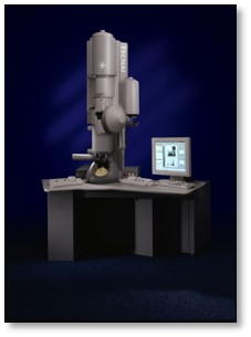 Model: Tecnai F30 / Image courtesy: FEI Company |
|---|---|
| Microscopy | Model: FEI TECNAI F30 |
| Electron gun | Electron source:  FIELD EMISSION GUN / Accelerating voltage: 300 kV / Illumination mode: FLOOD BEAM FIELD EMISSION GUN / Accelerating voltage: 300 kV / Illumination mode: FLOOD BEAM |
| Electron lens | Mode: BRIGHT FIELD / Nominal defocus max: 2000 nm / Nominal defocus min: 1500 nm |
| Image recording | Electron dose: 50 e/Å2 / Film or detector model: GATAN K3 (6k x 4k) |
- Processing
| EM software |
| ||||||||||||||||||||||||
|---|---|---|---|---|---|---|---|---|---|---|---|---|---|---|---|---|---|---|---|---|---|---|---|---|---|
| CTF correction | Type: PHASE FLIPPING AND AMPLITUDE CORRECTION | ||||||||||||||||||||||||
| 3D reconstruction | Resolution: 3.56 Å / Resolution method: FSC 0.143 CUT-OFF / Num. of particles: 125617 / Symmetry type: POINT | ||||||||||||||||||||||||
| Refinement | Highest resolution: 3.56 Å Stereochemistry target values: REAL-SPACE (WEIGHTED MAP SUM AT ATOM CENTERS) | ||||||||||||||||||||||||
| Refine LS restraints |
|
