 Movie
Movie Controller


+ Open data

- Basic information
| Entry |  | |||||||||
|---|---|---|---|---|---|---|---|---|---|---|
| Title | The structure of PldB-PA5088 complex state_1 | |||||||||
 Map data Map data | ||||||||||
 Sample Sample |
| |||||||||
 Keywords Keywords | Inhibitor / Complex / IMMUNE SYSTEM | |||||||||
| Function / homology |  Function and homology information Function and homology information | |||||||||
| Biological species |  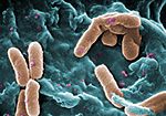 Pseudomonas aeruginosa (bacteria) Pseudomonas aeruginosa (bacteria) | |||||||||
| Method | single particle reconstruction / cryo EM / Resolution: 3.59 Å | |||||||||
 Authors Authors | Yang JW / Yang XY / Li ZQ | |||||||||
| Funding support |  China, 1 items China, 1 items
| |||||||||
 Citation Citation | Journal: Commun Biol / Year: 2026 Title: Structural basis of cooperative neutralization of the PldB toxin by immunity proteins in Pseudomonas aeruginosa. Authors: Xiaoyun Yang / Jiawen Yang / Hong Wang / Xiuhua Liu / Zongqiang Li / Abstract: PldB, a type VI secretion system-dependent phospholipase D (PLD) effector secreted by multidrug-resistant Pseudomonas aeruginosa, alters host cell membrane permeability and facilitates pathogen ...PldB, a type VI secretion system-dependent phospholipase D (PLD) effector secreted by multidrug-resistant Pseudomonas aeruginosa, alters host cell membrane permeability and facilitates pathogen internalization. Its cytotoxic activity is neutralized by three cognate immunity proteins-PA5086, PA5087, and PA5088-which protect the bacterium from self-intoxication. However, the underlying mechanism remains unclear. Through quantitative and qualitative analyses, we demonstrate that these three immunity proteins function cooperatively to inhibit PldB toxicity. Cryogenic electron microscopy of the PldB-PA5088 complex reveals that PA5088 binds to the HKD2 domain of PldB primarily through electrostatic interactions, markedly reducing the volume of its active center. Interaction studies using domain‑specific truncated PldB variants, together with enzyme activity assays, identify distinct copy numbers and binding regions for PA5086, PA5087, and PA5088 in their association with PldB. Collectively, our findings provide mechanistic insights into immunity protein-mediated neutralization of PldB toxicity, offering a potential foundation for designing PLD-targeting therapeutics against P. aeruginosa infection. | |||||||||
| History |
|
- Structure visualization
| Supplemental images |
|---|

- Downloads & links
-EMDB archive
| Map data | emd_67105.map.gz | 44.4 MB | EMDB map data format | |
|---|---|---|---|---|
| Header (meta data) | emd-67105-v30.xmlemd-67105.xml | 15.7 KB 15.7 KB | Display Display | EMDB header |
| Images |  emd_67105.png emd_67105.png | 48.6 KB | ||
| Filedesc metadata | emd-67105.cif.gz | 6 KB | ||
| Others | emd_67105_half_map_1.map.gzemd_67105_half_map_2.map.gz | 84.5 MB 84.5 MB | ||
| Archive directory |  https://data.pdbj.org/pub/emdb/structures/EMD-67105ftp://data.pdbj.org/pub/emdb/structures/EMD-67105 https://data.pdbj.org/pub/emdb/structures/EMD-67105ftp://data.pdbj.org/pub/emdb/structures/EMD-67105 | HTTPS FTP |
-Related structure data
| Related structure data |  9xq9MC  9xqmC M: atomic model generated by this map C: citing same article ( |
|---|---|
| Similar structure data |

-Links
| EMDB pages | EMDB (EBI/PDBe) / EMDataResource |
|---|
-Map
| File | Download / File: emd_67105.map.gz / Format: CCP4 / Size: 91.1 MB / Type: IMAGE STORED AS FLOATING POINT NUMBER (4 BYTES) | ||||||||||||||||||||||||||||||||||||
|---|---|---|---|---|---|---|---|---|---|---|---|---|---|---|---|---|---|---|---|---|---|---|---|---|---|---|---|---|---|---|---|---|---|---|---|---|---|
| Projections & slices | Image control
Images are generated by Spider. | ||||||||||||||||||||||||||||||||||||
| Voxel size | X=Y=Z: 0.668 Å | ||||||||||||||||||||||||||||||||||||
| Density |
| ||||||||||||||||||||||||||||||||||||
| Symmetry | Space group: 1 | ||||||||||||||||||||||||||||||||||||
| Details | EMDB XML:
|
 Z (Sec.)
 Y (Row.)
 X (Col.)


-Supplemental data
-Half map: #2
| File | emd_67105_half_map_1.map | ||||||||||||
|---|---|---|---|---|---|---|---|---|---|---|---|---|---|
| Projections & Slices |
| ||||||||||||
| Density Histograms |


-Half map: #1
| File | emd_67105_half_map_2.map | ||||||||||||
|---|---|---|---|---|---|---|---|---|---|---|---|---|---|
| Projections & Slices |
| ||||||||||||
| Density Histograms |


- Sample components
-Entire : Complex of Pldb-PA5088
| Entire | Name: Complex of Pldb-PA5088 |
|---|---|
| Components |
|
-Supramolecule #1: Complex of Pldb-PA5088
| Supramolecule | Name: Complex of Pldb-PA5088 / type: complex / ID: 1 / Parent: 0 / Macromolecule list: all |
|---|---|
| Source (natural) | Organism: Pseudomonas aeruginosa (bacteria) |
-Macromolecule #1: PLD phosphodiesterase domain-containing protein
| Macromolecule | Name: PLD phosphodiesterase domain-containing protein / type: protein_or_peptide / ID: 1 / Number of copies: 1 / Enantiomer: LEVO |
|---|---|
| Source (natural) | Organism: Pseudomonas aeruginosa (bacteria) |
| Molecular weight | Theoretical: 80.928703 KDa |
| Recombinant expression | Organism:  Escherichia coli (E. coli) Escherichia coli (E. coli) |
| Sequence | String: TLDWFANKAF YPPRAGVHIK PLINGQAAFD AVHAAMEAAR HSIDIITWGF DPAMRFKRPD GPRIGELLQT KGREGVQARV LVWSNQLAR LKENTIPGAG VGGSGGTWAG SGVASGSAVD NEVLRLEQRR QHNLNLIARQ QEALERSERL HREGRLPSFD P RGAAHARA ...String: TLDWFANKAF YPPRAGVHIK PLINGQAAFD AVHAAMEAAR HSIDIITWGF DPAMRFKRPD GPRIGELLQT KGREGVQARV LVWSNQLAR LKENTIPGAG VGGSGGTWAG SGVASGSAVD NEVLRLEQRR QHNLNLIARQ QEALERSERL HREGRLPSFD P RGAAHARA RIAELEAENA EIQRTLDSSE AQGYGGKRGS GGTRQDPWGQ IFTRDWFKAV RGGGLQNVEF RTRDFEQTAR PV MNGEQVR LVNGRLQSLI HLLRADGNDD LGIGQLLVLT QFASHHQKMV LVDYGSPQAI GFVMGHNMHR NYWDTSAHLF DDR AAGRDP GFGPWQDISM QVQGPVLADL SRNFSEAWDL ETPWYKRWFS TPSLTAERDA LPLPKIATPA SNSVAQICRT QPQD DERSI LEHYLKALGN ATDYVYMENQ YFRYAGFAER LRKTAQVRKA RGVPGDLYLF VVTNTPDSSD ASKTTYDMMK GLGQE QLMP QVQRDLAHDL REKREQLKQV RENLHPDPYV RRGQENNIER LERKIEALEE KGVTPEVEQR LGDLGAQEIP GLAKNT GED DKPYQLAEAP GLKVVVATLA TSDPAPGSPP PARLSAEAEA ALGAPPLKAR YKHIYVHSKL LLVDDLYTLL SSANINV RS MHGDSELGIA QPNPDLARAM REELWGAHVG RLAETTEKNF ELWNQKMDDN WKAQVADEPF TSHLLRFWDV TTPYSQNL T VD UniProtKB: PLD phosphodiesterase domain-containing protein |
-Macromolecule #2: Sel1 repeat family protein
| Macromolecule | Name: Sel1 repeat family protein / type: protein_or_peptide / ID: 2 / Number of copies: 1 / Enantiomer: LEVO |
|---|---|
| Source (natural) | Organism: Pseudomonas aeruginosa (bacteria) |
| Molecular weight | Theoretical: 30.509408 KDa |
| Recombinant expression | Organism: Escherichia coli (E. coli) |
| Sequence | String: AELRPFICVN EKDHLPSLDP QADAWYREAV ALAKPDTLRP WDRIVDLYSK AVERGHWKAM HNLASLYRTG WPGGVEKDTQ KALDLYQKM IDLKVPQGFY DMAAMIGNRA GVKNPATDGL TFLDKAASLG NPPALTELGR LYIYVAGQDE LGLKYTNCAA G QGYAPANY ...String: AELRPFICVN EKDHLPSLDP QADAWYREAV ALAKPDTLRP WDRIVDLYSK AVERGHWKAM HNLASLYRTG WPGGVEKDTQ KALDLYQKM IDLKVPQGFY DMAAMIGNRA GVKNPATDGL TFLDKAASLG NPPALTELGR LYIYVAGQDE LGLKYTNCAA G QGYAPANY ELAMYYRLVA HNYPKAAGYY LLAASQGNDD AAFFMSGVFD KTSPDVDRMW YAPDEKLHKL YDGIYDQLAA DP DLRFPNL IKDHPLPPHP TQGYDADRPD WKPGQ UniProtKB: Sel1 repeat family protein |
-Experimental details
-Structure determination
| Method | cryo EM |
|---|---|
 Processing Processing | single particle reconstruction |
| Aggregation state | particle |
-Sample preparation
| Buffer | pH: 8 |
|---|---|
| Vitrification | Cryogen name: ETHANE |
- Electron microscopy
| Microscope | FEI TECNAI F30 |
|---|---|
| Image recording | Film or detector model: GATAN K3 (6k x 4k) / Average electron dose: 50.0 e/Å2 |
| Electron beam | Acceleration voltage: 300 kV / Electron source:  FIELD EMISSION GUN FIELD EMISSION GUN |
| Electron optics | Illumination mode: FLOOD BEAM / Imaging mode: BRIGHT FIELD / Nominal defocus max: 2.0 µm / Nominal defocus min: 1.5 µm |
| Experimental equipment | 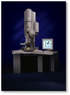 Model: Tecnai F30 / Image courtesy: FEI Company |
