 Movie
Movie Controller

[English] 日本語
 Yorodumi
- PDB-9xgx: Cryo-EM structure of SARS-CoV-2 receptor binding domain in comple... -
+ Open data

- Basic information
| Entry | Database: PDB / ID: 9xgx | |||||||||
|---|---|---|---|---|---|---|---|---|---|---|
| Title | Cryo-EM structure of SARS-CoV-2 receptor binding domain in complex with SR-23 Fab (local refinement of RBD and Fv) | |||||||||
 Components Components |
| |||||||||
 Keywords Keywords | VIRAL PROTEIN/IMMUNE SYSTEM / Viral protein / antibody / VIRAL PROTEIN-IMMUNE SYSTEM complex | |||||||||
| Function / homology |  Function and homology information Function and homology informationsymbiont-mediated disruption of host tissue / Maturation of spike protein / host cell surface / Translation of Structural Proteins / Virion Assembly and Release / Lectin pathway of complement activation / host extracellular region / symbiont-mediated-mediated suppression of host tetherin activity / structural constituent of virion / Induction of Cell-Cell Fusion ...symbiont-mediated disruption of host tissue / Maturation of spike protein / host cell surface / Translation of Structural Proteins / Virion Assembly and Release / Lectin pathway of complement activation / host extracellular region / symbiont-mediated-mediated suppression of host tetherin activity / structural constituent of virion / Induction of Cell-Cell Fusion / positive regulation of viral entry into host cell / Initial triggering of complement / membrane fusion / host cell endoplasmic reticulum-Golgi intermediate compartment membrane / Attachment and Entry / entry receptor-mediated virion attachment to host cell / receptor-mediated virion attachment to host cell / host cell surface receptor binding / symbiont-mediated suppression of host innate immune response / endocytosis involved in viral entry into host cell / receptor ligand activity / fusion of virus membrane with host plasma membrane / fusion of virus membrane with host endosome membrane / viral envelope / symbiont entry into host cell / virion attachment to host cell / host cell plasma membrane / SARS-CoV-2 activates/modulates innate and adaptive immune responses / virion membrane / membrane / identical protein binding / plasma membrane Similarity search - Function | |||||||||
| Biological species |  Homo sapiens (human) Homo sapiens (human) 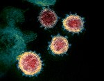 Severe acute respiratory syndrome coronavirus 2 Severe acute respiratory syndrome coronavirus 2 | |||||||||
| Method | ELECTRON MICROSCOPY / single particle reconstruction / cryo EM / Resolution: 2.8 Å | |||||||||
 Authors Authors | Kim, U.J. / Wang, D.M. / Yoon, G.Y. / Cho, H.S. | |||||||||
| Funding support |  Korea, Republic Of, 2items Korea, Republic Of, 2items
| |||||||||
 Citation Citation | Journal: J Med Virol / Year: 2026 Title: Broad Neutralizing Activity of Monoclonal Antibodies Against the Omicron Variants Isolated From Patients With Early Severe Acute Respiratory Syndrome Coronavirus-2. Authors: Da Sol Kim / Uijin Kim / Hye-Min Woo / Hansaem Lee / Eun-Seong Jo / Min Jeong Noh / So-Young Lee / Byoung Kwon Park / Jeong-Sun Yang / Kyung-Chang Kim / Joo-Yeon Lee / Dong-Min Wang / Hyun- ...Authors: Da Sol Kim / Uijin Kim / Hye-Min Woo / Hansaem Lee / Eun-Seong Jo / Min Jeong Noh / So-Young Lee / Byoung Kwon Park / Jeong-Sun Yang / Kyung-Chang Kim / Joo-Yeon Lee / Dong-Min Wang / Hyun-Soo Cho / Hyun-Joo Kim / Abstract: Since its emergence in 2019, severe acute respiratory syndrome coronavirus-2 (SARS-CoV-2) has caused a global pandemic driven by rapid mutation and high transmissibility. Current therapeutic ...Since its emergence in 2019, severe acute respiratory syndrome coronavirus-2 (SARS-CoV-2) has caused a global pandemic driven by rapid mutation and high transmissibility. Current therapeutic strategies may not effectively address the continuously evolving mutant strains, underscoring the need for treatments with broad neutralizing activity. Two monoclonal antibodies (mAbs): SR-23 and CS-42, isolated from convalescent patients infected with SARS-CoV-2 during the early phase of the 2020 pandemic, demonstrated significant neutralizing activities against a variety of variants. Specifically, SR-23 exhibited neutralizing activity against BA.5 and XBB1.5, whereas CS-42 showed efficacy against Delta, BA.1, and BA.2 variants. In addition, combining the two mAbs demonstrated improved neutralization activity. Cryo-electron microscopy (cryo-EM) structural analysis revealed that SR-23 and CS-42 bind to nonoverlapping epitopes on the SARS-CoV-2 spike protein. Furthermore, both antibodies exhibited strong therapeutic effects in K18-hACE2 mice infected with D614G, BA.2, and XBB.1.5. These findings provide insights into the development of antibody-based therapeutics targeting emerging Omicron variants and suggest that combined administration could broaden neutralizing activity. | |||||||||
| History |
|
- Structure visualization
| Structure viewer | Molecule: MolmilJmol/JSmol |
|---|
- Downloads & links
-Download
| PDBx/mmCIF format | 9xgx.cif.gz | 162.6 KB | Display | PDBx/mmCIF format |
|---|---|---|---|---|
| PDB format | pdb9xgx.ent.gz | 128.6 KB | Display | PDB format |
| PDBx/mmJSON format | 9xgx.json.gz | Tree view | PDBx/mmJSON format | |
| Others |  Other downloads Other downloads |
-Validation report
| Arichive directory | https://data.pdbj.org/pub/pdb/validation_reports/xg/9xgxftp://data.pdbj.org/pub/pdb/validation_reports/xg/9xgx | HTTPS FTP |
|---|
-Related structure data
| Related structure data |  66861MC  9xgwC M: map data used to model this data C: citing same article ( |
|---|---|
| Similar structure data |

-Links
 PDBj
PDBj


- Assembly
| Deposited unit | 
|
|---|---|
| 1 |
|
-Components
| #1: Antibody | Mass: 12577.061 Da / Num. of mol.: 1 Source method: isolated from a genetically manipulated source Source: (gene. exp.) Homo sapiens (human) / Cell line (production host): HEK293 / Production host: Homo sapiens (human) |
|---|---|
| #2: Antibody | Mass: 11810.103 Da / Num. of mol.: 1 Source method: isolated from a genetically manipulated source Source: (gene. exp.) Homo sapiens (human) / Cell line (production host): HEK293 / Production host: Homo sapiens (human) |
| #3: Protein | Mass: 22320.039 Da / Num. of mol.: 1 Source method: isolated from a genetically manipulated source Details: Sample sequence starts from 333 Source: (gene. exp.) Severe acute respiratory syndrome coronavirus 2Gene: S, 2 / Production host:   Spodoptera frugiperda (fall armyworm) / References: UniProt: P0DTC2 Spodoptera frugiperda (fall armyworm) / References: UniProt: P0DTC2 |
| #4: Sugar | ChemComp-NAG /   Type: D-saccharide, beta linking / Mass: 221.208 Da / Num. of mol.: 1 / Source method: obtained synthetically / Formula: C8H15NO6 Type: D-saccharide, beta linking / Mass: 221.208 Da / Num. of mol.: 1 / Source method: obtained synthetically / Formula: C8H15NO6 |
| Has ligand of interest | N |
| Has protein modification | Y |
-Experimental details
-Experiment
| Experiment | Method: ELECTRON MICROSCOPY |
|---|---|
| EM experiment | Aggregation state: PARTICLE / 3D reconstruction method: single particle reconstruction |
- Sample preparation
| Component |
| ||||||||||||||||||||||||
|---|---|---|---|---|---|---|---|---|---|---|---|---|---|---|---|---|---|---|---|---|---|---|---|---|---|
| Molecular weight |
| ||||||||||||||||||||||||
| Source (natural) |
| ||||||||||||||||||||||||
| Source (recombinant) |
| ||||||||||||||||||||||||
| Buffer solution | pH: 7.5 / Details: 20mM Tris-HCl, 150mM NaCl | ||||||||||||||||||||||||
| Buffer component |
| ||||||||||||||||||||||||
| Specimen | Conc.: 0.05 mg/ml / Embedding applied: NO / Shadowing applied: NO / Staining applied: NO / Vitrification applied: YES | ||||||||||||||||||||||||
| Specimen support | Grid material: COPPER / Grid mesh size: 200 divisions/in. / Grid type: Quantifoil R1.2/1.3 | ||||||||||||||||||||||||
| Vitrification | Instrument: FEI VITROBOT MARK IV / Cryogen name: ETHANE / Humidity: 100 % / Chamber temperature: 277.15 K |
- Electron microscopy imaging
| Experimental equipment |  Model: Titan Krios / Image courtesy: FEI Company |
|---|---|
| Microscopy | Model: TFS KRIOS |
| Electron gun | Electron source:  FIELD EMISSION GUN / Accelerating voltage: 300 kV / Illumination mode: FLOOD BEAM FIELD EMISSION GUN / Accelerating voltage: 300 kV / Illumination mode: FLOOD BEAM |
| Electron lens | Mode: BRIGHT FIELD / Calibrated magnification: 105000 X / Nominal defocus max: 1700 nm / Nominal defocus min: 700 nm |
| Specimen holder | Cryogen: NITROGEN / Specimen holder model: FEI TITAN KRIOS AUTOGRID HOLDER |
| Image recording | Electron dose: 67.6 e/Å2 / Film or detector model: GATAN K3 BIOCONTINUUM (6k x 4k) |
- Processing
| EM software |
| |||||||||||||||||||||||||||||||||||
|---|---|---|---|---|---|---|---|---|---|---|---|---|---|---|---|---|---|---|---|---|---|---|---|---|---|---|---|---|---|---|---|---|---|---|---|---|
| CTF correction | Type: PHASE FLIPPING AND AMPLITUDE CORRECTION | |||||||||||||||||||||||||||||||||||
| Particle selection | Num. of particles selected: 2200000 | |||||||||||||||||||||||||||||||||||
| 3D reconstruction | Resolution: 2.8 Å / Resolution method: FSC 0.143 CUT-OFF / Num. of particles: 115041 / Algorithm: FOURIER SPACE / Num. of class averages: 1 / Symmetry type: POINT | |||||||||||||||||||||||||||||||||||
| Atomic model building | Protocol: RIGID BODY FIT / Space: REAL | |||||||||||||||||||||||||||||||||||
| Atomic model building | Source name: AlphaFold / Type: in silico model | |||||||||||||||||||||||||||||||||||
| Refinement | Highest resolution: 2.8 Å Stereochemistry target values: REAL-SPACE (WEIGHTED MAP SUM AT ATOM CENTERS) | |||||||||||||||||||||||||||||||||||
| Refine LS restraints |
|
