 Movie
Movie Controller

[English] 日本語
 Yorodumi
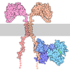
- PDB-9tg8: Drebrin actin binding domain 1 conformation A (ABD1a) bound to F-actin -
+ Open data

- Basic information
| Entry | Database: PDB / ID: 9tg8 | |||||||||||||||||||||||||||
|---|---|---|---|---|---|---|---|---|---|---|---|---|---|---|---|---|---|---|---|---|---|---|---|---|---|---|---|---|
| Title | Drebrin actin binding domain 1 conformation A (ABD1a) bound to F-actin | |||||||||||||||||||||||||||
 Components Components |
| |||||||||||||||||||||||||||
 Keywords Keywords | STRUCTURAL PROTEIN / F-actin binding protein | |||||||||||||||||||||||||||
| Function / homology |  Function and homology information Function and homology informationpositive regulation of receptor localization to synapse / regulation of dendrite development / profilin binding / positive regulation of dendritic spine morphogenesis / actomyosin / positive regulation of synaptic plasticity / gap junction / RHOD GTPase cycle / neural precursor cell proliferation / RHOBTB1 GTPase cycle ...positive regulation of receptor localization to synapse / regulation of dendrite development / profilin binding / positive regulation of dendritic spine morphogenesis / actomyosin / positive regulation of synaptic plasticity / gap junction / RHOD GTPase cycle / neural precursor cell proliferation / RHOBTB1 GTPase cycle / cytoskeletal motor activator activity / generation of neurons / myosin heavy chain binding / tropomyosin binding / actin filament bundle / troponin I binding / filamentous actin / mesenchyme migration / cortical cytoskeleton / regulation of neuronal synaptic plasticity / skeletal muscle myofibril / striated muscle thin filament / RHOH GTPase cycle / actin filament bundle assembly / skeletal muscle thin filament assembly / actin monomer binding / postsynaptic cytosol / RHOBTB2 GTPase cycle / skeletal muscle fiber development / actin filament polymerization / stress fiber / titin binding / actin filament organization / filopodium / actin filament / protein sequestering activity / Hydrolases; Acting on acid anhydrides; Acting on acid anhydrides to facilitate cellular and subcellular movement / calcium-dependent protein binding / actin cytoskeleton / lamellipodium / growth cone / actin binding / cell body / cytoskeleton / postsynaptic membrane / postsynaptic density / cadherin binding / protein domain specific binding / hydrolase activity / positive regulation of gene expression / calcium ion binding / dendrite / glutamatergic synapse / magnesium ion binding / ATP binding / identical protein binding / cytoplasm Similarity search - Function | |||||||||||||||||||||||||||
| Biological species |  Homo sapiens (human) Homo sapiens (human)  Oryctolagus cuniculus (rabbit) Oryctolagus cuniculus (rabbit) | |||||||||||||||||||||||||||
| Method | ELECTRON MICROSCOPY / single particle reconstruction / cryo EM / Resolution: 2.37 Å | |||||||||||||||||||||||||||
 Authors Authors | Zhao, W. / Abis, G. / Oozeer, F. / Mulvaney, T. / Nagar, N. / Topf, M. / Gordon-Weeks, P.R. / Conte, M.R. / Atherton, J. | |||||||||||||||||||||||||||
| Funding support |  United Kingdom, 8items United Kingdom, 8items
| |||||||||||||||||||||||||||
 Citation Citation | Journal: Nat Commun / Year: 2026 Title: Structural mechanisms of drebrin-mediated F-actin network modulation. Authors: W Zhao / L Y Chu / G Abis / F Oozeer / T Mulvaney / N Nagar / M Topf / P R Gordon-Weeks / M R Conte / J Atherton /  Abstract: Drebrin modulates F-actin networks and links them to other intracellular components, regulating crucial processes including neuritogenesis, synaptic plasticity, virus internalisation and cancer ...Drebrin modulates F-actin networks and links them to other intracellular components, regulating crucial processes including neuritogenesis, synaptic plasticity, virus internalisation and cancer invasion. Using single-particle cryo-EM we characterise drebrin's interaction with F-actin through two separate conserved actin binding domains (ABD1 and ABD2), revealing structural bases for its F-actin-modulating properties. We describe a multimodal interaction where drebrin's ABD1 can adopt two conformations and a long flexible loop connecting to ABD2 allows the two ABDs to occupy multiple relative positions along F-actin. The flexible loop connecting the two ABDs also confers some propensity to loosely bundle F-actin. Drebrin's ABDs bind across multiple actin protomers and their subdomains and modify the longitudinal inter-protomer interface, explaining its F-actin stabilising properties. Furthermore, we show drebrin's binding site on F-actin is shared with other critical actin-binding and regulatory proteins, explaining their competitive displacement. | |||||||||||||||||||||||||||
| History |
|
- Structure visualization
| Structure viewer | Molecule: MolmilJmol/JSmol |
|---|
- Downloads & links
-Download
| PDBx/mmCIF format | 9tg8.cif.gz | 186.2 KB | Display | PDBx/mmCIF format |
|---|---|---|---|---|
| PDB format | pdb9tg8.ent.gz | 142.9 KB | Display | PDB format |
| PDBx/mmJSON format | 9tg8.json.gz | Tree view | PDBx/mmJSON format | |
| Others |  Other downloads Other downloads |
-Validation report
| Arichive directory | https://data.pdbj.org/pub/pdb/validation_reports/tg/9tg8ftp://data.pdbj.org/pub/pdb/validation_reports/tg/9tg8 | HTTPS FTP |
|---|
-Related structure data
| Related structure data |  55893MC  9tg9C  9tgaC M: map data used to model this data C: citing same article ( |
|---|---|
| Similar structure data |


-Links
 PDBj
PDBj



- Assembly
| Deposited unit | 
|
|---|---|
| 1 |
|
-Components
| #1: Protein | Mass: 26188.834 Da / Num. of mol.: 1 Source method: isolated from a genetically manipulated source Source: (gene. exp.) Homo sapiens (human) / Gene: DBN1, D0S117E / Production host:   Escherichia coli BL21(DE3) (bacteria) / References: UniProt: Q16643 Escherichia coli BL21(DE3) (bacteria) / References: UniProt: Q16643 | ||||||||||
|---|---|---|---|---|---|---|---|---|---|---|---|
| #2: Protein | Mass: 42109.973 Da / Num. of mol.: 2 / Source method: isolated from a natural source / Source: (natural) Oryctolagus cuniculus (rabbit) / References: UniProt: P68135#3: Chemical |  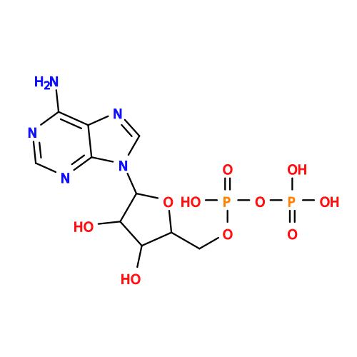 Mass: 427.201 Da / Num. of mol.: 2 / Source method: obtained synthetically / Formula: C10H15N5O10P2 / Comment: ADP, energy-carrying molecule*YM Mass: 427.201 Da / Num. of mol.: 2 / Source method: obtained synthetically / Formula: C10H15N5O10P2 / Comment: ADP, energy-carrying molecule*YM#4: Chemical |   Mass: 24.305 Da / Num. of mol.: 2 / Source method: obtained synthetically / Formula: Mg Mass: 24.305 Da / Num. of mol.: 2 / Source method: obtained synthetically / Formula: Mg#5: Water | ChemComp-HOH / |  Mass: 18.015 Da / Num. of mol.: 265 / Source method: isolated from a natural source / Formula: H2O Mass: 18.015 Da / Num. of mol.: 265 / Source method: isolated from a natural source / Formula: H2OHas ligand of interest | N | Has protein modification | Y | |
-Experimental details
-Experiment
| Experiment | Method: ELECTRON MICROSCOPY |
|---|---|
| EM experiment | Aggregation state: FILAMENT / 3D reconstruction method: single particle reconstruction |
- Sample preparation
| Component |
| ||||||||||||||||||||||||||||||
|---|---|---|---|---|---|---|---|---|---|---|---|---|---|---|---|---|---|---|---|---|---|---|---|---|---|---|---|---|---|---|---|
| Source (natural) |
| ||||||||||||||||||||||||||||||
| Source (recombinant) |
| ||||||||||||||||||||||||||||||
| Buffer solution | pH: 7.5 | ||||||||||||||||||||||||||||||
| Buffer component |
| ||||||||||||||||||||||||||||||
| Specimen | Embedding applied: NO / Shadowing applied: NO / Staining applied: NO / Vitrification applied: YES | ||||||||||||||||||||||||||||||
| Specimen support | Grid material: GOLD / Grid type: Au-flat 1.2/1.3 | ||||||||||||||||||||||||||||||
| Vitrification | Cryogen name: ETHANE |
- Electron microscopy imaging
| Experimental equipment |  Model: Titan Krios / Image courtesy: FEI Company |
|---|---|
| Microscopy | Model: TFS KRIOS |
| Electron gun | Electron source:  FIELD EMISSION GUN / Accelerating voltage: 300 kV / Illumination mode: FLOOD BEAM FIELD EMISSION GUN / Accelerating voltage: 300 kV / Illumination mode: FLOOD BEAM |
| Electron lens | Mode: BRIGHT FIELD / Nominal defocus max: 2500 nm / Nominal defocus min: 700 nm / C2 aperture diameter: 50 µm |
| Image recording | Electron dose: 45 e/Å2 / Film or detector model: TFS FALCON 4i (4k x 4k) |
- Processing
| EM software |
| ||||||||||||||||||||||||||||||||||||||||
|---|---|---|---|---|---|---|---|---|---|---|---|---|---|---|---|---|---|---|---|---|---|---|---|---|---|---|---|---|---|---|---|---|---|---|---|---|---|---|---|---|---|
| CTF correction | Type: PHASE FLIPPING AND AMPLITUDE CORRECTION | ||||||||||||||||||||||||||||||||||||||||
| 3D reconstruction | Resolution: 2.37 Å / Resolution method: FSC 0.143 CUT-OFF / Num. of particles: 102155 / Symmetry type: POINT | ||||||||||||||||||||||||||||||||||||||||
| Atomic model building | Protocol: OTHER | ||||||||||||||||||||||||||||||||||||||||
| Refinement | Highest resolution: 2.37 Å Stereochemistry target values: REAL-SPACE (WEIGHTED MAP SUM AT ATOM CENTERS) | ||||||||||||||||||||||||||||||||||||||||
| Refine LS restraints |
|
