 Movie
Movie Controller

[English] 日本語
 Yorodumi
- PDB-9s34: Structure of the Pyrococcus abyssi 20S proteasome alpha subunit b... -
+ Open data

- Basic information
| Entry | Database: PDB / ID: 9s34 | |||||||||||||||
|---|---|---|---|---|---|---|---|---|---|---|---|---|---|---|---|---|
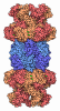
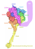
| Title | Structure of the Pyrococcus abyssi 20S proteasome alpha subunit bound to the capping protein APA1 | |||||||||||||||
 Components Components |
| |||||||||||||||
 Keywords Keywords | PROTEIN BINDING / Complex Proteasome Archaea Proteasome activator | |||||||||||||||
| Function / homology |  Function and homology information Function and homology informationthreonine-type endopeptidase activity / proteasome core complex, alpha-subunit complex / proteasomal protein catabolic process / ubiquitin-dependent protein catabolic process / cytoplasm Similarity search - Function | |||||||||||||||
| Biological species |   Pyrococcus abyssi (archaea) Pyrococcus abyssi (archaea) | |||||||||||||||
| Method | ELECTRON MICROSCOPY / single particle reconstruction / cryo EM / Resolution: 3.15 Å | |||||||||||||||
 Authors Authors | Musso, F. / Marino Puertas, L. / Weis, F. / Schoehn, G. / Franzetti, B. | |||||||||||||||
| Funding support |  France, 1items France, 1items
| |||||||||||||||
 Citation Citation | Journal: To Be Published Title: Structure of the Pyrococcus abyssi 20S proteasome alpha subunit bound to the capping protein APA1 Authors: Musso, F. / Marino Puertas, L. / Girard, E. / Gabel, F. / Coute, Y. / Flament, D. / Chenavier, F. / Weis, F. / Schoehn, G. / Franzetti, B. | |||||||||||||||
| History |
|
- Structure visualization
| Structure viewer | Molecule: MolmilJmol/JSmol |
|---|
- Downloads & links
-Download
| PDBx/mmCIF format | 9s34.cif.gz | 695.2 KB | Display | PDBx/mmCIF format |
|---|---|---|---|---|
| PDB format | pdb9s34.ent.gz | 582.6 KB | Display | PDB format |
| PDBx/mmJSON format | 9s34.json.gz | Tree view | PDBx/mmJSON format | |
| Others |  Other downloads Other downloads |
-Validation report
| Arichive directory | https://data.pdbj.org/pub/pdb/validation_reports/s3/9s34ftp://data.pdbj.org/pub/pdb/validation_reports/s3/9s34 | HTTPS FTP |
|---|
-Related structure data
| Related structure data |  54521MC C: citing same article ( M: map data used to model this data |
|---|---|
| Similar structure data |


-Links
 PDBj
PDBj

- Assembly
| Deposited unit | 
|
|---|---|
| 1 |
|
-Components
| #1: Protein | Mass: 33512.344 Da / Num. of mol.: 7 Source method: isolated from a genetically manipulated source Source: (gene. exp.) Pyrococcus abyssi (archaea) / Gene: PAB1363 / Production host:   Escherichia coli (E. coli) / References: UniProt: Q9UYJ3 Escherichia coli (E. coli) / References: UniProt: Q9UYJ3#2: Protein | Mass: 29035.293 Da / Num. of mol.: 7 Source method: isolated from a genetically manipulated source Source: (gene. exp.) Pyrococcus abyssi (archaea) / Gene: psmA, PYRAB06070, PAB0417 / Production host: Escherichia coli (E. coli) / References: UniProt: Q9V122Has protein modification | N | |
|---|
-Experimental details
-Experiment
| Experiment | Method: ELECTRON MICROSCOPY |
|---|---|
| EM experiment | Aggregation state: PARTICLE / 3D reconstruction method: single particle reconstruction |
- Sample preparation
| Component |
| ||||||||||||||||||||||||||||
|---|---|---|---|---|---|---|---|---|---|---|---|---|---|---|---|---|---|---|---|---|---|---|---|---|---|---|---|---|---|
| Molecular weight |
| ||||||||||||||||||||||||||||
| Source (natural) |
| ||||||||||||||||||||||||||||
| Source (recombinant) |
| ||||||||||||||||||||||||||||
| Buffer solution | pH: 8 Details: 50 mM Tris pH 8.0, 150 mM NaCl, 150 mM KCl, 10 mM MgCl2 | ||||||||||||||||||||||||||||
| Specimen | Embedding applied: NO / Shadowing applied: NO / Staining applied: NO / Vitrification applied: YES / Details: SEC-pure form of the 20S | ||||||||||||||||||||||||||||
| Vitrification | Instrument: FEI VITROBOT MARK IV / Cryogen name: ETHANE-PROPANE / Humidity: 100 % / Chamber temperature: 298 K |
- Electron microscopy imaging
| Microscopy | Model: TFS GLACIOS |
|---|---|
| Electron gun | Electron source:  FIELD EMISSION GUN / Accelerating voltage: 200 kV / Illumination mode: FLOOD BEAM FIELD EMISSION GUN / Accelerating voltage: 200 kV / Illumination mode: FLOOD BEAM |
| Electron lens | Mode: BRIGHT FIELD / Nominal defocus max: 2500 nm / Nominal defocus min: 1000 nm / Cs: 2.7 mm |
| Specimen holder | Cryogen: NITROGEN |
| Image recording | Electron dose: 40 e/Å2 / Detector mode: COUNTING / Film or detector model: GATAN K2 SUMMIT (4k x 4k) / Num. of grids imaged: 1 / Num. of real images: 4835 |
- Processing
| EM software |
| ||||||||||||||||||||||||||||||||
|---|---|---|---|---|---|---|---|---|---|---|---|---|---|---|---|---|---|---|---|---|---|---|---|---|---|---|---|---|---|---|---|---|---|
| CTF correction | Type: PHASE FLIPPING AND AMPLITUDE CORRECTION | ||||||||||||||||||||||||||||||||
| Particle selection | Num. of particles selected: 534489 | ||||||||||||||||||||||||||||||||
| Symmetry | Point symmetry: C7 (7 fold cyclic) | ||||||||||||||||||||||||||||||||
| 3D reconstruction | Resolution: 3.15 Å / Resolution method: FSC 0.143 CUT-OFF / Num. of particles: 50092 Details: 50,092 particles aligning to the complex were used for Non Uniform refinement with an imposed C7 symmetry. This yielded the consensus alignment at 3.23 Angstrom, which served as a base for ...Details: 50,092 particles aligning to the complex were used for Non Uniform refinement with an imposed C7 symmetry. This yielded the consensus alignment at 3.23 Angstrom, which served as a base for the focused refinements used to make this composite map. Upon reconstruction with imposed C7 symmetry, the two proteasome Beta1-2 subunits of the 20S became merged together. We noticed that the same was happening when the 20S proteasome was reconstructed without an imposed symmetry, suggesting a random distribution of the two subunit types in the Beta rings. Due to the high degree of similarity between the two proteins (>70 percent sequence identity) and the small number of particles, we opted for not continuing with further 3D classification. Symmetry type: POINT | ||||||||||||||||||||||||||||||||
| Atomic model building | Protocol: RIGID BODY FIT / Space: REAL / Target criteria: Cross correlation Details: Model building done in coot, refinement in Phenix. No models built for the merged Beta 1-2 chains of the 20S proteasome, due to the identical folding of the two proteins and the very small ...Details: Model building done in coot, refinement in Phenix. No models built for the merged Beta 1-2 chains of the 20S proteasome, due to the identical folding of the two proteins and the very small (single side chains) differences between the two subunits, which prevented effective 3D classification with the small number of particles left. IMPORTANT: the outlier Y204 nonplanar bond was modelled as such based on a higher resolution X-ray model, to be deposited soon. | ||||||||||||||||||||||||||||||||
| Atomic model building |
|
