 Movie
Movie Controller

[English] 日本語
 Yorodumi
- EMDB-54521: Structure of the Pyrococcus abyssi 20S proteasome bound to the ar... -
+ Open data

- Basic information
| Entry |  | |||||||||
|---|---|---|---|---|---|---|---|---|---|---|
| Title | Structure of the Pyrococcus abyssi 20S proteasome bound to the archaeal activator APA1 | |||||||||
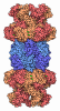
 Map data Map data | Ensemble view of the 20S proteasome from Pyrococcus abyssi and the capping protein Q9UYJ3 (APA1) | |||||||||
 Sample Sample |
| |||||||||
 Keywords Keywords | Complex Proteasome Archaea Proteasome activator / PROTEIN BINDING | |||||||||
| Function / homology |  Function and homology information Function and homology informationthreonine-type endopeptidase activity / proteasome core complex, alpha-subunit complex / proteasomal protein catabolic process / ubiquitin-dependent protein catabolic process / cytoplasm Similarity search - Function | |||||||||
| Biological species |   Pyrococcus abyssi (archaea) Pyrococcus abyssi (archaea) | |||||||||
| Method | single particle reconstruction / cryo EM / Resolution: 3.15 Å | |||||||||
 Authors Authors | Musso F / Marino Puertas L / Weis F / Schoehn G / Franzetti B | |||||||||
| Funding support |  France, 1 items France, 1 items
| |||||||||
 Citation Citation | Journal: To Be Published Title: Structure of the Pyrococcus abyssi 20S proteasome alpha subunit bound to the capping protein APA1 Authors: Musso F / Marino Puertas L / Girard E / Gabel F / Coute Y / Flament D / Chenavier F / Weis F / Schoehn G / Franzetti B | |||||||||
| History |
|
- Structure visualization
| Supplemental images |
|---|

- Downloads & links
-EMDB archive
| Map data | emd_54521.map.gz | 396.3 MB | EMDB map data format | |
|---|---|---|---|---|
| Header (meta data) | emd-54521-v30.xmlemd-54521.xml | 20.4 KB 20.4 KB | Display Display | EMDB header |
| Images |  emd_54521.png emd_54521.png | 161.7 KB | ||
| Filedesc metadata | emd-54521.cif.gz | 7.2 KB | ||
| Archive directory |  https://data.pdbj.org/pub/emdb/structures/EMD-54521ftp://data.pdbj.org/pub/emdb/structures/EMD-54521 https://data.pdbj.org/pub/emdb/structures/EMD-54521ftp://data.pdbj.org/pub/emdb/structures/EMD-54521 | HTTPS FTP |
-Related structure data
| Related structure data |  9s34MC C: citing same article ( M: atomic model generated by this map |
|---|---|
| Similar structure data |


-Links
| EMDB pages | EMDB (EBI/PDBe) / EMDataResource |
|---|---|
| Related items in Molecule of the Month |


-Map
| File | Download / File: emd_54521.map.gz / Format: CCP4 / Size: 421.9 MB / Type: IMAGE STORED AS FLOATING POINT NUMBER (4 BYTES) | ||||||||||||||||||||||||||||||||||||
|---|---|---|---|---|---|---|---|---|---|---|---|---|---|---|---|---|---|---|---|---|---|---|---|---|---|---|---|---|---|---|---|---|---|---|---|---|---|
| Annotation | Ensemble view of the 20S proteasome from Pyrococcus abyssi and the capping protein Q9UYJ3 (APA1) | ||||||||||||||||||||||||||||||||||||
| Projections & slices | Image control
Images are generated by Spider. | ||||||||||||||||||||||||||||||||||||
| Voxel size | X=Y=Z: 1.145 Å | ||||||||||||||||||||||||||||||||||||
| Density |
| ||||||||||||||||||||||||||||||||||||
| Symmetry | Space group: 1 | ||||||||||||||||||||||||||||||||||||
| Details | EMDB XML:
|
 Z (Sec.)
 Y (Row.)
 X (Col.)


-Supplemental data
- Sample components
-Entire : 20S Proteasome from P. abyssi in complex with the heptameric prot...
| Entire | Name: 20S Proteasome from P. abyssi in complex with the heptameric protein APA1 (Q9UYJ3) |
|---|---|
| Components |
|
-Supramolecule #1: 20S Proteasome from P. abyssi in complex with the heptameric prot...
| Supramolecule | Name: 20S Proteasome from P. abyssi in complex with the heptameric protein APA1 (Q9UYJ3) type: cell / ID: 1 / Parent: 0 / Macromolecule list: all Details: Assembled 20S core particle reconstituted in vitro from recombinant expression of its three subunit types: alpha, beta-1, beta-2. Beta 2 subunit bears an N-terminal His-tag. Capping protein ...Details: Assembled 20S core particle reconstituted in vitro from recombinant expression of its three subunit types: alpha, beta-1, beta-2. Beta 2 subunit bears an N-terminal His-tag. Capping protein APA1 sitting on the outer Alpha ring. |
|---|---|
| Source (natural) | Organism: Pyrococcus abyssi (archaea) |
-Supramolecule #2: 20S Proteasome from P. abyssi
| Supramolecule | Name: 20S Proteasome from P. abyssi / type: complex / ID: 2 / Parent: 1 / Macromolecule list: #2 |
|---|---|
| Source (natural) | Organism: Pyrococcus abyssi (archaea) |
| Molecular weight | Theoretical: 711 KDa |
-Supramolecule #3: Archaeal proteasome activator 1
| Supramolecule | Name: Archaeal proteasome activator 1 / type: complex / ID: 3 / Parent: 1 / Macromolecule list: #1 Details: Heptameric form of the protein, in complex with the proteasome. No tags. |
|---|---|
| Source (natural) | Organism: Pyrococcus abyssi (archaea) |
| Molecular weight | Theoretical: 234 KDa |
-Macromolecule #1: Archaeal proteasome activator 1
| Macromolecule | Name: Archaeal proteasome activator 1 / type: protein_or_peptide / ID: 1 / Number of copies: 7 / Enantiomer: LEVO |
|---|---|
| Source (natural) | Organism: Pyrococcus abyssi (archaea) |
| Molecular weight | Theoretical: 33.512344 KDa |
| Recombinant expression | Organism:   Escherichia coli (E. coli) Escherichia coli (E. coli) |
| Sequence | String: MKASKLAVKL LKTENDEIIY YDPIYHGRTL KVIGIDDDPA LVMEYLLAQY KEKGYNVIVF DTSGKFPTSL FDNILRIEEN SPAGLDPLK LARVGLIKDP YSAVTIIQTI YELDRASTEK LYADFIKGKV NSMDEVVRSK ESYAEVINES YTELDEMLFS G EPMKVPES ...String: MKASKLAVKL LKTENDEIIY YDPIYHGRTL KVIGIDDDPA LVMEYLLAQY KEKGYNVIVF DTSGKFPTSL FDNILRIEEN SPAGLDPLK LARVGLIKDP YSAVTIIQTI YELDRASTEK LYADFIKGKV NSMDEVVRSK ESYAEVINES YTELDEMLFS G EPMKVPES CLIDLSALNS ITLTGNAFLI LAAMLEDRRS VAYGLYDVSV LTFTDSGNAG LPLITRAARK RVSIVGTRYA LD QILNIPG PTLLLYNDPD VQSAIYESQG VPQGFRRFVE KGEGAYIVRS PETIEVEFGM LLR UniProtKB: Uncharacterized protein |
-Macromolecule #2: Proteasome subunit alpha
| Macromolecule | Name: Proteasome subunit alpha / type: protein_or_peptide / ID: 2 / Number of copies: 7 / Enantiomer: LEVO |
|---|---|
| Source (natural) | Organism: Pyrococcus abyssi (archaea) |
| Molecular weight | Theoretical: 29.035293 KDa |
| Recombinant expression | Organism: Escherichia coli (E. coli) |
| Sequence | String: MAFVPPQAGY DRAITVFSPD GRLFQVNYAR EAVKRGATAV GVKCKDGVVL AVEKRITSRL IEPESYEKIF QIDDHIAAAS SGIIADARV LVNRARLEAQ IHRLTYGEPA PLAVIVKKIC DLKQMHTQYG GVRPFGAALL MAGVNDKPEL YETDPSGAYF A WKAVAIGS ...String: MAFVPPQAGY DRAITVFSPD GRLFQVNYAR EAVKRGATAV GVKCKDGVVL AVEKRITSRL IEPESYEKIF QIDDHIAAAS SGIIADARV LVNRARLEAQ IHRLTYGEPA PLAVIVKKIC DLKQMHTQYG GVRPFGAALL MAGVNDKPEL YETDPSGAYF A WKAVAIGS GRNTAMAIFE DKYRDDMTLD EAIKLAIFAL AKTMEKPSAE NIEVAVITVK DKKFRKLSKE EIEKFLGEVM KE VEEEEVK EKEEDYSELD SHY UniProtKB: Proteasome subunit alpha |
-Experimental details
-Structure determination
| Method | cryo EM |
|---|---|
 Processing Processing | single particle reconstruction |
| Aggregation state | particle |
-Sample preparation
| Buffer | pH: 8 Details: 50 mM Tris pH 8.0, 150 mM NaCl, 150 mM KCl, 10 mM MgCl2 |
|---|---|
| Vitrification | Cryogen name: ETHANE-PROPANE / Chamber humidity: 100 % / Chamber temperature: 298 K / Instrument: FEI VITROBOT MARK IV |
| Details | SEC-pure form of the 20S |
- Electron microscopy
| Microscope | TFS GLACIOS |
|---|---|
| Image recording | Film or detector model: GATAN K2 SUMMIT (4k x 4k) / Detector mode: COUNTING / Number grids imaged: 1 / Number real images: 4835 / Average electron dose: 40.0 e/Å2 |
| Electron beam | Acceleration voltage: 200 kV / Electron source:  FIELD EMISSION GUN FIELD EMISSION GUN |
| Electron optics | Illumination mode: FLOOD BEAM / Imaging mode: BRIGHT FIELD / Cs: 2.7 mm / Nominal defocus max: 2.5 µm / Nominal defocus min: 1.0 µm |
| Sample stage | Cooling holder cryogen: NITROGEN |
+Image processing
-Atomic model buiding 1
| Initial model |
| |||||||||
|---|---|---|---|---|---|---|---|---|---|---|
| Details | Model building done in coot, refinement in Phenix. No models built for the merged Beta 1-2 chains of the 20S proteasome, due to the identical folding of the two proteins and the very small (single side chains) differences between the two subunits, which prevented effective 3D classification with the small number of particles left. IMPORTANT: the outlier Y204 nonplanar bond was modelled as such based on a higher resolution X-ray model, to be deposited soon. | |||||||||
| Refinement | Space: REAL / Protocol: RIGID BODY FIT / Target criteria: Cross correlation | |||||||||
| Output model | PDB-9s34: |

