 Movie
Movie Controller


+ Open data

- Basic information
| Entry | Database: PDB / ID: 9ox7 | |||||||||||||||||||||||||||||||||
|---|---|---|---|---|---|---|---|---|---|---|---|---|---|---|---|---|---|---|---|---|---|---|---|---|---|---|---|---|---|---|---|---|---|---|
| Title | In situ microtubule structure in the axon of a human neuron | |||||||||||||||||||||||||||||||||
 Components Components |
| |||||||||||||||||||||||||||||||||
 Keywords Keywords | STRUCTURAL PROTEIN / Human in-situ microtubule / axon / cytoskeleton | |||||||||||||||||||||||||||||||||
| Function / homology |  Function and homology information Function and homology informationpyramidal neuron differentiation / glial cell differentiation / Post-chaperonin tubulin folding pathway / cytoskeleton-dependent intracellular transport / Cargo trafficking to the periciliary membrane / dentate gyrus development / Carboxyterminal post-translational modifications of tubulin / Microtubule-dependent trafficking of connexons from Golgi to the plasma membrane / axonemal microtubule / organelle transport along microtubule ...pyramidal neuron differentiation / glial cell differentiation / Post-chaperonin tubulin folding pathway / cytoskeleton-dependent intracellular transport / Cargo trafficking to the periciliary membrane / dentate gyrus development / Carboxyterminal post-translational modifications of tubulin / Microtubule-dependent trafficking of connexons from Golgi to the plasma membrane / axonemal microtubule / organelle transport along microtubule / forebrain morphogenesis / cerebellar cortex morphogenesis / Sealing of the nuclear envelope (NE) by ESCRT-III / Intraflagellar transport / neuron projection arborization / Formation of tubulin folding intermediates by CCT/TriC / Gap junction assembly / smoothened signaling pathway / homeostasis of number of cells within a tissue / motor behavior / Kinesins / positive regulation of axon guidance / Prefoldin mediated transfer of substrate to CCT/TriC / response to L-glutamate / Assembly and cell surface presentation of NMDA receptors / COPI-independent Golgi-to-ER retrograde traffic / centrosome cycle / sperm principal piece / COPI-dependent Golgi-to-ER retrograde traffic / startle response / 'de novo' protein folding / intercellular bridge / flagellated sperm motility / regulation of synapse organization / sperm end piece / locomotory exploration behavior / Recycling pathway of L1 / microtubule polymerization / ciliary tip / response to tumor necrosis factor / response to mechanical stimulus / neuron apoptotic process / sperm flagellum / adult locomotory behavior / RHO GTPases activate IQGAPs / microtubule-based process / Hedgehog 'off' state / COPI-mediated anterograde transport / Activation of AMPK downstream of NMDARs / condensed chromosome / Loss of Nlp from mitotic centrosomes / Loss of proteins required for interphase microtubule organization from the centrosome / Recruitment of mitotic centrosome proteins and complexes / MHC class II antigen presentation / cellular response to calcium ion / Recruitment of NuMA to mitotic centrosomes / Anchoring of the basal body to the plasma membrane / visual learning / HSP90 chaperone cycle for steroid hormone receptors (SHR) in the presence of ligand / Mitotic Prometaphase / EML4 and NUDC in mitotic spindle formation / AURKA Activation by TPX2 / Resolution of Sister Chromatid Cohesion / Translocation of SLC2A4 (GLUT4) to the plasma membrane / neuromuscular junction / neuron migration / memory / cerebral cortex development / RHO GTPases Activate Formins / intracellular protein transport / synapse organization / microtubule cytoskeleton organization / PKR-mediated signaling / recycling endosome / mitotic spindle / modulation of chemical synaptic transmission / Schaffer collateral - CA1 synapse / structural constituent of cytoskeleton / cytoplasmic ribonucleoprotein granule / HCMV Early Events / microtubule cytoskeleton / Aggrephagy / The role of GTSE1 in G2/M progression after G2 checkpoint / cilium / Separation of Sister Chromatids / Regulation of PLK1 Activity at G2/M Transition / neuron differentiation / mitotic cell cycle / protein-folding chaperone binding / microtubule binding / microtubule / Hydrolases; Acting on acid anhydrides; Acting on GTP to facilitate cellular and subcellular movement / protein stabilization / protein heterodimerization activity / cell division / hydrolase activity / GTPase activity / GTP binding / protein-containing complex binding / structural molecule activity Similarity search - Function | |||||||||||||||||||||||||||||||||
| Biological species |  Homo sapiens (human) Homo sapiens (human) | |||||||||||||||||||||||||||||||||
| Method | ELECTRON MICROSCOPY / helical reconstruction / cryo EM / Resolution: 2.69 Å | |||||||||||||||||||||||||||||||||
 Authors Authors | Zehr, E.A. / Sun, S. / Roll-Mecak, A. | |||||||||||||||||||||||||||||||||
| Funding support |  United States, 1items United States, 1items
| |||||||||||||||||||||||||||||||||
 Citation Citation | Journal: Nat Struct Mol Biol / Year: 2026 Title: Microtubules in the axon are GDP bound but adopt a stable GTP-like expanded state. Authors: Elena A Zehr / Shufeng Sun / Stephanie L Sarbanes / Antonina Roll-Mecak / Abstract: Microtubules scaffold cells, supporting signaling and cargo transport. They assemble from GTP-tubulin, which hydrolyzes to GDP-tubulin during polymerization. GTP-microtubule lattices are stable; GDP ...Microtubules scaffold cells, supporting signaling and cargo transport. They assemble from GTP-tubulin, which hydrolyzes to GDP-tubulin during polymerization. GTP-microtubule lattices are stable; GDP lattices depolymerize rapidly. In vitro, hydrolysis triggers lattice compaction. Lattice spacing regulates motors and microtubule-associated proteins; however, the conformation of tubulin in microtubules in cells is unknown. Here, we present the atomic-resolution cryo-electron microscopy structure of human microtubules in situ, in the axons of human cortical neurons derived from induced pluripotent stem cells (iPS cells). Our 2.7-Å-resolution reconstruction delineates bound water molecules and reveals that axonal microtubules adopt an expanded GTP-like lattice, despite being GDP bound. Using cryo-electron tomography and power spectrum analysis, we find that, unlike in axons, microtubules in undifferentiated iPS cells are compacted. Therefore, lattice expansion is part of neuronal differentiation. Our work provides molecular insights into neurogenesis and has implications for understanding microtubule stability and effector recruitment in neurons. | |||||||||||||||||||||||||||||||||
| History |
|
- Structure visualization
| Structure viewer | Molecule: MolmilJmol/JSmol |
|---|
- Downloads & links
-Download
| PDBx/mmCIF format | 9ox7.cif.gz | 658.7 KB | Display | PDBx/mmCIF format |
|---|---|---|---|---|
| PDB format | pdb9ox7.ent.gz | 542.3 KB | Display | PDB format |
| PDBx/mmJSON format | 9ox7.json.gz | Tree view | PDBx/mmJSON format | |
| Others |  Other downloads Other downloads |
-Validation report
| Arichive directory | https://data.pdbj.org/pub/pdb/validation_reports/ox/9ox7ftp://data.pdbj.org/pub/pdb/validation_reports/ox/9ox7 | HTTPS FTP |
|---|
-Related structure data
| Related structure data |  70956MC M: map data used to model this data C: citing same article ( |
|---|---|
| Similar structure data | |
| Experimental dataset #1 | Data reference: 10.6019/EMPIAR-13239 / Db source: EMPIAR Details: Cryo-electron tomography of microtubules in axons from human cortical neurons derived from induced pluripotent stem cells (iPSCs) |
| Experimental dataset #2 | Data reference: 10.6019/EMPIAR-13240 / Db source: EMPIAR Details: Cryo-electron tomography of microtubules in undifferentiated induced pluripotent stem cells (iPSCs) |
| Experimental dataset #3 | Data reference: 10.6019/EMPIAR-13241 / Db source: EMPIAR Details: Cryo-electron tomography of in vitro assembled, GDP-bound, porcine brain microtubules |
| Experimental dataset #4 | Data reference: 10.6019/EMPIAR-13244 / Db source: EMPIAR Details: Cryo-electron tomography of in vitro assembled, GMPCPP-bound, porcine brain microtubules |
| Experimental dataset #5 | Data reference: 10.6019/EMPIAR-13243 / Db source: EMPIAR Details: Cryo-electron tomography of in vitro assembled, GMPCPP-bound, porcine brain microtubules |
| Experimental dataset #6 | Data reference: 10.6019/EMPIAR-13242 / Db source: EMPIAR Details: Cryo-electron tomography of in vitro assembled, GDP-bound, porcine brain microtubules |
-Links
 PDBj
PDBj










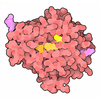

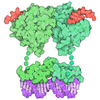
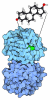

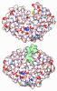
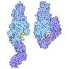
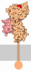
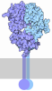
- Assembly
| Deposited unit | 
|
|---|---|
| 1 |
|
-Components
-Protein , 2 types, 8 molecules BDEFACGH
| #1: Protein | Mass: 49999.887 Da / Num. of mol.: 4 / Source method: isolated from a natural source / Source: (natural) Homo sapiens (human) / Cell line: WTC11 / Tissue: Neuronal / References: UniProt: Q9BVA1#2: Protein | Mass: 50188.441 Da / Num. of mol.: 4 / Source method: isolated from a natural source / Source: (natural) Homo sapiens (human) / Cell line: WTC11 / Tissue: Neuronal / References: UniProt: Q71U36 |
|---|
-Non-polymers , 4 types, 44 molecules 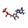

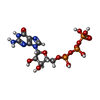

| #3: Chemical | ChemComp-GDP / 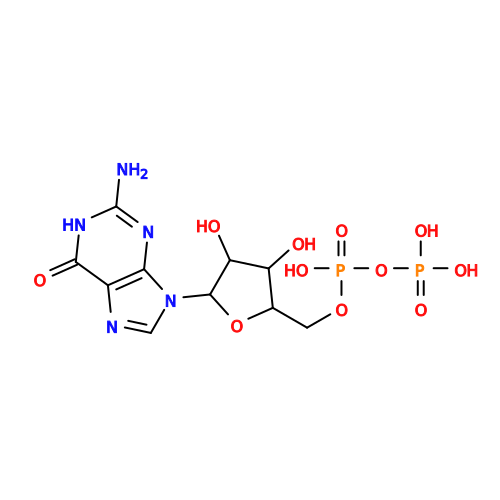 Type: RNA linking / Mass: 443.201 Da / Num. of mol.: 4 / Source method: obtained synthetically / Formula: C10H15N5O11P2 / Feature type: SUBJECT OF INVESTIGATION / Comment: GDP, energy-carrying molecule*YM Type: RNA linking / Mass: 443.201 Da / Num. of mol.: 4 / Source method: obtained synthetically / Formula: C10H15N5O11P2 / Feature type: SUBJECT OF INVESTIGATION / Comment: GDP, energy-carrying molecule*YM#4: Chemical | ChemComp-MG /  Mass: 24.305 Da / Num. of mol.: 4 / Source method: obtained synthetically / Formula: Mg Mass: 24.305 Da / Num. of mol.: 4 / Source method: obtained synthetically / Formula: Mg#5: Chemical | ChemComp-GTP / 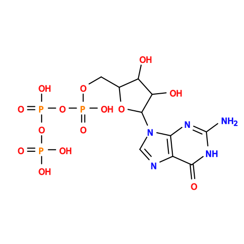 Mass: 523.180 Da / Num. of mol.: 4 / Source method: obtained synthetically / Formula: C10H16N5O14P3 / Comment: GTP, energy-carrying molecule*YM Mass: 523.180 Da / Num. of mol.: 4 / Source method: obtained synthetically / Formula: C10H16N5O14P3 / Comment: GTP, energy-carrying molecule*YM#6: Water | ChemComp-HOH / | Mass: 18.015 Da / Num. of mol.: 32 / Source method: isolated from a natural source / Formula: H2O |
|---|
-Details
| Has ligand of interest | Y |
|---|---|
| Has protein modification | N |
-Experimental details
-Experiment
| Experiment | Method: ELECTRON MICROSCOPY |
|---|---|
| EM experiment | Aggregation state: FILAMENT / 3D reconstruction method: helical reconstruction |
- Sample preparation
| Component | Name: Axonal alpha1A and betaIIB microtubule / Type: COMPLEX / Entity ID: #1-#2 / Source: NATURAL |
|---|---|
| Molecular weight | Value: 1 MDa / Experimental value: NO |
| Source (natural) | Organism: Homo sapiens (human) / Cellular location: axon / Tissue: neuronal |
| Buffer solution | pH: 7.2 |
| Specimen | Conc.: 0.55 mg/ml / Embedding applied: NO / Shadowing applied: NO / Staining applied: NO / Vitrification applied: YES |
| Specimen support | Grid material: GOLD / Grid mesh size: 200 divisions/in. / Grid type: Quantifoil R2/2 |
| Vitrification | Instrument: FEI VITROBOT MARK II / Cryogen name: ETHANE / Humidity: 85 % / Chamber temperature: 303 K |
- Electron microscopy imaging
| Experimental equipment |  Model: Titan Krios / Image courtesy: FEI Company |
|---|---|
| Microscopy | Model: TFS KRIOS |
| Electron gun | Electron source:  FIELD EMISSION GUN / Accelerating voltage: 300 kV / Illumination mode: FLOOD BEAM FIELD EMISSION GUN / Accelerating voltage: 300 kV / Illumination mode: FLOOD BEAM |
| Electron lens | Mode: BRIGHT FIELD / Nominal magnification: 105000 X / Nominal defocus max: 2500 nm / Nominal defocus min: 1200 nm / Cs: 2.7 mm / C2 aperture diameter: 70 µm / Alignment procedure: COMA FREE |
| Image recording | Average exposure time: 2.03 sec. / Electron dose: 56.57 e/Å2 / Film or detector model: GATAN K3 (6k x 4k) / Num. of grids imaged: 6 / Num. of real images: 3017 |
| EM imaging optics | Energyfilter name: GIF Bioquantum / Energyfilter slit width: 20 eV |
- Processing
| EM software |
| ||||||||||||||||||||||||||||||||||||||||
|---|---|---|---|---|---|---|---|---|---|---|---|---|---|---|---|---|---|---|---|---|---|---|---|---|---|---|---|---|---|---|---|---|---|---|---|---|---|---|---|---|---|
| CTF correction | Type: PHASE FLIPPING AND AMPLITUDE CORRECTION | ||||||||||||||||||||||||||||||||||||||||
| Helical symmerty | Angular rotation/subunit: -27.6723 ° / Axial rise/subunit: 9.60742 Å / Axial symmetry: C1 | ||||||||||||||||||||||||||||||||||||||||
| Particle selection | Num. of particles selected: 288841 | ||||||||||||||||||||||||||||||||||||||||
| 3D reconstruction | Resolution: 2.69 Å / Resolution method: FSC 0.143 CUT-OFF / Num. of particles: 1887666 / Algorithm: FOURIER SPACE / Symmetry type: HELICAL | ||||||||||||||||||||||||||||||||||||||||
| Atomic model building | B value: 69 / Protocol: FLEXIBLE FIT / Space: REAL / Target criteria: cross-correlation | ||||||||||||||||||||||||||||||||||||||||
| Atomic model building | PDB-ID: 8v4k Accession code: 8v4k / Source name: PDB / Type: experimental model |

