Movie
Movie Controller

[English] 日本語
 Yorodumi
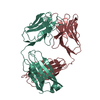
- PDB-9me7: Antibody fragments from mAb21 and mAb824 bound to the adhesin pro... -
+ Open data

- Basic information
| Entry | Database: PDB / ID: 9me7 | |||||||||||||||
|---|---|---|---|---|---|---|---|---|---|---|---|---|---|---|---|---|
| Title | Antibody fragments from mAb21 and mAb824 bound to the adhesin protein FimH containing alpha-methyl mannose | |||||||||||||||
 Components Components |
| |||||||||||||||
 Keywords Keywords | CELL ADHESION/IMMUNE SYSTEM / Fimbrial tip / Lectin domain / Antibody fragments / Antibody-target complex / CELL ADHESION / CELL ADHESION-IMMUNE SYSTEM complex | |||||||||||||||
| Function / homology |  Function and homology information Function and homology informationpilus tip / mechanosensory behavior / Attachment of bacteria to epithelial cells / cell adhesion involved in single-species biofilm formation / pilus / cell-substrate adhesion / D-mannose binding / host cell membrane / cell adhesion Similarity search - Function | |||||||||||||||
| Biological species |   | |||||||||||||||
| Method | ELECTRON MICROSCOPY / single particle reconstruction / cryo EM / Resolution: 3.1 Å | |||||||||||||||
 Authors Authors | Hvorecny, K.L. / Magala, P. / Klevit, R.E. / Kollman, J.M. | |||||||||||||||
| Funding support |  United States, 4items United States, 4items
| |||||||||||||||
 Citation Citation | Journal: Nat Commun / Year: 2025 Title: Antibodies disrupt bacterial adhesion by ligand mimicry and allosteric interference. Authors: Kelli L Hvorecny / Gianluca Interlandi / Tim S Veth / Pavel Aprikian / Anna Manchenko / Veronika L Tchesnokova / Miles S Dickinson / Joel D Quispe / Nicholas M Riley / Rachel E Klevit / ...Authors: Kelli L Hvorecny / Gianluca Interlandi / Tim S Veth / Pavel Aprikian / Anna Manchenko / Veronika L Tchesnokova / Miles S Dickinson / Joel D Quispe / Nicholas M Riley / Rachel E Klevit / Pearl Magala / Evgeni V Sokurenko / Justin M Kollman / Abstract: A critical step in infections is the attachment of microorganisms to host cells using lectins that bind glycans, making lectins promising antimicrobial targets. Upon binding mannosylated glycans, ...A critical step in infections is the attachment of microorganisms to host cells using lectins that bind glycans, making lectins promising antimicrobial targets. Upon binding mannosylated glycans, FimH, an adhesin in E. coli, undergoes an allosteric transition from an inactive to an active conformation that can act as a catch-bond. Distinct monoclonal antibodies that alter FimH glycan binding are available, but the mechanisms of action remain unclear. Here, we use cryo-electron microscopy, mass spectrometry, adhesion assays, and molecular dynamics simulations to determine the structure-function relationships underlying antibody-FimH binding. Our study demonstrates four mechanisms of action: ligand mimicry by an N-linked, high-mannose glycan; stabilization of the ligand pocket in the inactive state; conformational trapping of the active and inactive states; and locking of the ligand pocket through long-range allosteric effects. These structures reveal multiple mechanisms of antibody responses to an allosteric protein and provide blueprints for antimicrobials that target adhesins. | |||||||||||||||
| History |
|
- Structure visualization
| Structure viewer | Molecule: MolmilJmol/JSmol |
|---|
- Downloads & links
-Download
| PDBx/mmCIF format | 9me7.cif.gz | 211.6 KB | Display | PDBx/mmCIF format |
|---|---|---|---|---|
| PDB format | pdb9me7.ent.gz | 150.3 KB | Display | PDB format |
| PDBx/mmJSON format | 9me7.json.gz | Tree view | PDBx/mmJSON format | |
| Others |  Other downloads Other downloads |
-Validation report
| Arichive directory | https://data.pdbj.org/pub/pdb/validation_reports/me/9me7ftp://data.pdbj.org/pub/pdb/validation_reports/me/9me7 | HTTPS FTP |
|---|
-Related structure data
| Related structure data |  48186MC  9me4C  9me5C  9me6C  9ptmC M: map data used to model this data C: citing same article ( |
|---|---|
| Similar structure data |


-Links
 PDBj
PDBj

- Assembly
| Deposited unit | 
|
|---|---|
| 1 |
|
-Components
-Antibody , 4 types, 4 molecules IJMN
| #2: Antibody | Mass: 22526.971 Da / Num. of mol.: 1 / Source method: isolated from a natural source / Source: (natural) |
|---|---|
| #3: Antibody | Mass: 21143.547 Da / Num. of mol.: 1 / Source method: isolated from a natural source / Source: (natural) |
| #4: Antibody | Mass: 21757.852 Da / Num. of mol.: 1 / Source method: isolated from a natural source / Source: (natural) |
| #5: Antibody | Mass: 19737.736 Da / Num. of mol.: 1 / Source method: isolated from a natural source / Source: (natural) |
-Protein / Sugars , 2 types, 2 molecules A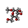
| #1: Protein | Mass: 31488.260 Da / Num. of mol.: 1 Source method: isolated from a genetically manipulated source Source: (gene. exp.) |
|---|---|
| #6: Sugar | ChemComp-MMA /  Type: D-saccharide / Mass: 194.182 Da / Num. of mol.: 1 / Source method: obtained synthetically / Formula: C7H14O6 / Feature type: SUBJECT OF INVESTIGATION Type: D-saccharide / Mass: 194.182 Da / Num. of mol.: 1 / Source method: obtained synthetically / Formula: C7H14O6 / Feature type: SUBJECT OF INVESTIGATION |
-Details
| Has ligand of interest | Y |
|---|---|
| Has protein modification | Y |
-Experimental details
-Experiment
| Experiment | Method: ELECTRON MICROSCOPY |
|---|---|
| EM experiment | Aggregation state: PARTICLE / 3D reconstruction method: single particle reconstruction |
- Sample preparation
| Component |
| ||||||||||||||||||||||||
|---|---|---|---|---|---|---|---|---|---|---|---|---|---|---|---|---|---|---|---|---|---|---|---|---|---|
| Molecular weight |
| ||||||||||||||||||||||||
| Source (natural) |
| ||||||||||||||||||||||||
| Source (recombinant) | Organism: | ||||||||||||||||||||||||
| Buffer solution | pH: 7.4 | ||||||||||||||||||||||||
| Specimen | Embedding applied: NO / Shadowing applied: NO / Staining applied: NO / Vitrification applied: YES | ||||||||||||||||||||||||
| Specimen support | Grid material: GOLD / Grid mesh size: 300 divisions/in. / Grid type: Quantifoil R2/1 | ||||||||||||||||||||||||
| Vitrification | Cryogen name: ETHANE |
- Electron microscopy imaging
| Experimental equipment |  Model: Titan Krios / Image courtesy: FEI Company |
|---|---|
| Microscopy | Model: TFS KRIOS |
| Electron gun | Electron source:  FIELD EMISSION GUN / Accelerating voltage: 300 kV / Illumination mode: FLOOD BEAM FIELD EMISSION GUN / Accelerating voltage: 300 kV / Illumination mode: FLOOD BEAM |
| Electron lens | Mode: BRIGHT FIELD / Nominal magnification: 105000 X / Nominal defocus max: 1500 nm / Nominal defocus min: 750 nm |
| Specimen holder | Cryogen: NITROGEN / Specimen holder model: FEI TITAN KRIOS AUTOGRID HOLDER |
| Image recording | Electron dose: 42 e/Å2 / Film or detector model: GATAN K3 BIOQUANTUM (6k x 4k) / Num. of grids imaged: 1 / Num. of real images: 7832 |
- Processing
| EM software | Name: PHENIX / Version: 1.21.1_5286 / Category: model refinement | |||||||||||||||||||||
|---|---|---|---|---|---|---|---|---|---|---|---|---|---|---|---|---|---|---|---|---|---|---|
| CTF correction | Type: PHASE FLIPPING AND AMPLITUDE CORRECTION | |||||||||||||||||||||
| 3D reconstruction | Resolution: 3.1 Å / Resolution method: FSC 0.143 CUT-OFF / Num. of particles: 163363 / Algorithm: FOURIER SPACE / Num. of class averages: 1 / Symmetry type: POINT | |||||||||||||||||||||
| Atomic model building | Protocol: FLEXIBLE FIT / Space: REAL | |||||||||||||||||||||
| Atomic model building |
| |||||||||||||||||||||
| Refinement | Cross valid method: NONE |