 Movie
Movie Controller

[English] 日本語
 Yorodumi
- PDB-9ibw: Dark-state structure of human short-wave-sensitive opsin (OPN1SW) -
+ Open data

- Basic information
| Entry | Database: PDB / ID: 9ibw | |||||||||||||||||||||||||||
|---|---|---|---|---|---|---|---|---|---|---|---|---|---|---|---|---|---|---|---|---|---|---|---|---|---|---|---|---|
| Title | Dark-state structure of human short-wave-sensitive opsin (OPN1SW) | |||||||||||||||||||||||||||
 Components Components |
| |||||||||||||||||||||||||||
 Keywords Keywords | MEMBRANE PROTEIN / human daylight vision / GPCR / OPN1SW / blue cone opsin | |||||||||||||||||||||||||||
| Function / homology |  Function and homology information Function and homology informationDefective visual phototransduction due to OPN1SW loss of function / cone photoreceptor disc membrane / The retinoid cycle in cones (daylight vision) / Opsins / absorption of visible light / G protein-coupled photoreceptor activity / cellular response to UV-A / cellular response to light stimulus / phototransduction / photoreceptor outer segment ...Defective visual phototransduction due to OPN1SW loss of function / cone photoreceptor disc membrane / The retinoid cycle in cones (daylight vision) / Opsins / absorption of visible light / G protein-coupled photoreceptor activity / cellular response to UV-A / cellular response to light stimulus / phototransduction / photoreceptor outer segment / visual perception / photoreceptor inner segment / electron transport chain / adenylate cyclase-inhibiting G protein-coupled receptor signaling pathway / photoreceptor disc membrane / signaling receptor activity / G alpha (i) signalling events / electron transfer activity / periplasmic space / iron ion binding / G protein-coupled receptor signaling pathway / heme binding / perinuclear region of cytoplasm / signal transduction / plasma membrane Similarity search - Function | |||||||||||||||||||||||||||
| Biological species |  Homo sapiens (human) Homo sapiens (human)  Escherichia coli (E. coli) Escherichia coli (E. coli)synthetic construct (others) | |||||||||||||||||||||||||||
| Method | ELECTRON MICROSCOPY / single particle reconstruction / cryo EM / Resolution: 3.22 Å | |||||||||||||||||||||||||||
 Authors Authors | Schmidt, S.L. / Isaikina, P. | |||||||||||||||||||||||||||
| Funding support | European Union, 1items
| |||||||||||||||||||||||||||
 Citation Citation | Journal: Science / Year: 2026 Title: Illuminating the molecular basis of human daylight vision. Authors: Sarah L Schmidt / Jakub Dostal / Saumik Sen / Andrej Hovan / Deborah Walter / Martin V Appleby / Asato Kojima / Hideaki E Kato / John H Beale / Miroslav Kloz / Gebhard F X Schertler / Polina Isaikina /    Abstract: Photopic vision, including fast motion and color perception in daylight, is mediated by cone opsins, specialized G protein-coupled receptors (GPCRs). Despite sharing the same chromophore, the three ...Photopic vision, including fast motion and color perception in daylight, is mediated by cone opsins, specialized G protein-coupled receptors (GPCRs). Despite sharing the same chromophore, the three receptor subtypes absorb light at different wavelengths of the visible spectrum. The molecular mechanisms governing their spectral properties and exceptionally rapid responses remain largely unknown. We report cryo-electron microscopy structures of the human blue-sensitive (OPN1SW) and green-sensitive (OPN1MW) cone opsins in their dark-adapted states, combined with femtosecond-resolution spectroscopy, functional assays, and advanced simulations. The data reveal distinct chromophore stabilization mechanisms across human visual opsins and specific sequence adaptations in the GPCR microswitch motifs, underlining their structural plasticity and distinct activation mechanisms. These findings delineate the molecular basis of the evolutionary refinements fulfilling the needs of vision in daylight. | |||||||||||||||||||||||||||
| History |
|
- Structure visualization
| Structure viewer | Molecule: MolmilJmol/JSmol |
|---|
- Downloads & links
-Download
| PDBx/mmCIF format | 9ibw.cif.gz | 144.3 KB | Display | PDBx/mmCIF format |
|---|---|---|---|---|
| PDB format | pdb9ibw.ent.gz | 105.8 KB | Display | PDB format |
| PDBx/mmJSON format | 9ibw.json.gz | Tree view | PDBx/mmJSON format | |
| Others |  Other downloads Other downloads |
-Validation report
| Arichive directory | https://data.pdbj.org/pub/pdb/validation_reports/ib/9ibwftp://data.pdbj.org/pub/pdb/validation_reports/ib/9ibw | HTTPS FTP |
|---|
-Related structure data
| Related structure data |  52814MC  9i3tC  9qp4C M: map data used to model this data C: citing same article ( |
|---|---|
| Similar structure data |


-Links
 PDBj
PDBj










- Assembly
| Deposited unit | 
|
|---|---|
| 1 |
|
-Components
| #1: Protein | Mass: 54494.750 Da / Num. of mol.: 1 Source method: isolated from a genetically manipulated source Source: (gene. exp.) Homo sapiens (human), (gene. exp.) Escherichia coli (E. coli)Gene: OPN1SW, BCP, cybC / Production host:   Spodoptera frugiperda (fall armyworm) / References: UniProt: P03999, UniProt: P0ABE7 Spodoptera frugiperda (fall armyworm) / References: UniProt: P03999, UniProt: P0ABE7 |
|---|---|
| #2: Antibody | Mass: 23812.252 Da / Num. of mol.: 1 Source method: isolated from a genetically manipulated source Source: (gene. exp.) synthetic construct (others) / Production host:   Cricetulus griseus (Chinese hamster) Cricetulus griseus (Chinese hamster) |
| #3: Antibody | Mass: 24033.934 Da / Num. of mol.: 1 Source method: isolated from a genetically manipulated source Source: (gene. exp.) synthetic construct (others) / Production host: Cricetulus griseus (Chinese hamster) |
| #4: Chemical | ChemComp-RET / 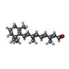 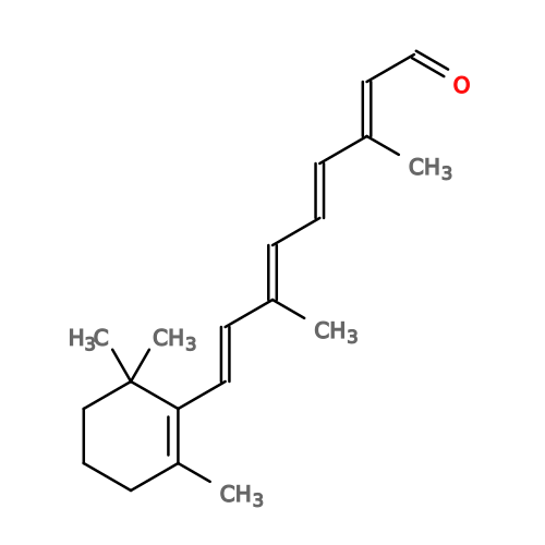 Mass: 284.436 Da / Num. of mol.: 1 / Source method: obtained synthetically / Formula: C20H28O / Feature type: SUBJECT OF INVESTIGATION Mass: 284.436 Da / Num. of mol.: 1 / Source method: obtained synthetically / Formula: C20H28O / Feature type: SUBJECT OF INVESTIGATION |
| Has ligand of interest | Y |
| Has protein modification | Y |
-Experimental details
-Experiment
| Experiment | Method: ELECTRON MICROSCOPY |
|---|---|
| EM experiment | Aggregation state: PARTICLE / 3D reconstruction method: single particle reconstruction |
- Sample preparation
| Component | Name: Human short-wave-sensitive opsin fused to soluble cytochrome b562 in complex with SPR2070 Fab Type: COMPLEX / Entity ID: #1-#3 / Source: RECOMBINANT |
|---|---|
| Molecular weight | Experimental value: NO |
| Source (natural) | Organism: Homo sapiens (human) |
| Source (recombinant) | Organism: Spodoptera frugiperda (fall armyworm) |
| Buffer solution | pH: 6.8 |
| Specimen | Embedding applied: NO / Shadowing applied: NO / Staining applied: NO / Vitrification applied: YES |
| Vitrification | Cryogen name: ETHANE-PROPANE |
- Electron microscopy imaging
| Experimental equipment |  Model: Titan Krios / Image courtesy: FEI Company |
|---|---|
| Microscopy | Model: TFS KRIOS |
| Electron gun | Electron source:  FIELD EMISSION GUN / Accelerating voltage: 300 kV / Illumination mode: OTHER FIELD EMISSION GUN / Accelerating voltage: 300 kV / Illumination mode: OTHER |
| Electron lens | Mode: OTHER / Nominal defocus max: 2500 nm / Nominal defocus min: 1200 nm |
| Image recording | Electron dose: 50 e/Å2 / Film or detector model: FEI FALCON IV (4k x 4k) |
- Processing
| EM software | Name: PHENIX / Version: 1.21.2_5419 / Category: model refinement | ||||||||||||||||||||||||
|---|---|---|---|---|---|---|---|---|---|---|---|---|---|---|---|---|---|---|---|---|---|---|---|---|---|
| CTF correction | Type: PHASE FLIPPING AND AMPLITUDE CORRECTION | ||||||||||||||||||||||||
| 3D reconstruction | Resolution: 3.22 Å / Resolution method: FSC 0.143 CUT-OFF / Num. of particles: 330659 / Symmetry type: POINT | ||||||||||||||||||||||||
| Refinement | Highest resolution: 3.22 Å Stereochemistry target values: REAL-SPACE (WEIGHTED MAP SUM AT ATOM CENTERS) | ||||||||||||||||||||||||
| Refine LS restraints |
|
