+ Open data
- Basic information
| Entry | Database: PDB / ID: 9goa | ||||||
|---|---|---|---|---|---|---|---|
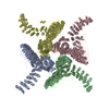
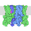
| Title | Pore state of alpha-Latrotoxin | ||||||
 Components Components | Alpha-latrotoxin-Lt1a | ||||||
 Keywords Keywords | TOXIN / Black widow spider toxin / Pore forming neurotoxin / Ankyrin repeat / presynaptic receptor activation | ||||||
| Function / homology |  Function and homology information Function and homology informationother organism cell membrane / host cell presynaptic membrane / exocytosis / toxin activity / extracellular region / membrane Similarity search - Function | ||||||
| Biological species |  Latrodectus tredecimguttatus (black widow) Latrodectus tredecimguttatus (black widow) | ||||||
| Method | ELECTRON MICROSCOPY / single particle reconstruction / cryo EM / Resolution: 3.2 Å | ||||||
 Authors Authors | Klink, B.U. / Gatsogiannis, C. / Kalyankumar, K.S. | ||||||
| Funding support |  Germany, 1items Germany, 1items
| ||||||
 Citation Citation |  Journal: Nat Commun / Year: 2024 Journal: Nat Commun / Year: 2024Title: Structural basis of α-latrotoxin transition to a cation-selective pore. Authors: B U Klink / A Alavizargar / K S Kalyankumar / M Chen / A Heuer / C Gatsogiannis /  Abstract: The potent neurotoxic venom of the black widow spider contains a cocktail of seven phylum-specific latrotoxins (LTXs), but only one, α-LTX, targets vertebrates. This 130 kDa toxin binds to ...The potent neurotoxic venom of the black widow spider contains a cocktail of seven phylum-specific latrotoxins (LTXs), but only one, α-LTX, targets vertebrates. This 130 kDa toxin binds to receptors at presynaptic nerve terminals and triggers a massive release of neurotransmitters. It is widely accepted that LTXs tetramerize and insert into the presynaptic membrane, thereby forming Ca-conductive pores, but the underlying mechanism remains poorly understood. LTXs are homologous and consist of an N-terminal region with three distinct domains, along with a C-terminal domain containing up to 22 consecutive ankyrin repeats. Here we report cryoEM structures of the vertebrate-specific α-LTX tetramer in its prepore and pore state. Our structures, in combination with AlphaFold2-based structural modeling and molecular dynamics simulations, reveal dramatic conformational changes in the N-terminal region of the complex. Four distinct helical bundles rearrange and together form a highly stable, 15 nm long, cation-impermeable coiled-coil stalk. This stalk, in turn, positions an N-terminal pair of helices within the membrane, thereby enabling the assembly of a cation-permeable channel. Taken together, these data give insight into a unique mechanism for membrane insertion and channel formation, characteristic of the LTX family, and provide the necessary framework for advancing novel therapeutics and biotechnological applications. #1:  Journal: BioRxiv / Year: 2024 Journal: BioRxiv / Year: 2024Title: Molecular mechanism of alpha-latrotoxin action Authors: Klink, B.U. / Alavizargar, A. / Kalyankumar, K.S. / Chen, M. / Heuer, A. / Gatsogiannis, C. | ||||||
| History |
|
- Structure visualization
| Structure viewer | Molecule:  Molmil Molmil Jmol/JSmol Jmol/JSmol |
|---|
- Downloads & links
- Download
| PDBx/mmCIF format |  9goa.cif.gz 9goa.cif.gz | 720 KB | Display |  PDBx/mmCIF format PDBx/mmCIF format |
|---|---|---|---|---|
| PDB format |  pdb9goa.ent.gz pdb9goa.ent.gz | 567.4 KB | Display |  PDB format PDB format |
| PDBx/mmJSON format |  9goa.json.gz 9goa.json.gz | Tree view |  PDBx/mmJSON format PDBx/mmJSON format | |
| Others |  Other downloads Other downloads |
-Validation report
| Arichive directory |  https://data.pdbj.org/pub/pdb/validation_reports/go/9goa https://data.pdbj.org/pub/pdb/validation_reports/go/9goa ftp://data.pdbj.org/pub/pdb/validation_reports/go/9goa ftp://data.pdbj.org/pub/pdb/validation_reports/go/9goa | HTTPS FTP |
|---|
-Related structure data
| Related structure data |  51495MC  9go9C C: citing same article ( M: map data used to model this data |
|---|---|
| Similar structure data | Similarity search - Function & homology  F&H Search F&H Search |
- Links
Links
- Assembly
| Deposited unit | 
|
|---|---|
| 1 |
|
- Components
| #1: Protein | Mass: 131749.141 Da / Num. of mol.: 4 / Source method: isolated from a natural source Details: alpha-latrotoxin activated via cleavage by Furin-like proteases Source: (natural)  Latrodectus tredecimguttatus (black widow) Latrodectus tredecimguttatus (black widow)Organ: venom gland / References: UniProt: P23631 Has protein modification | N | |
|---|
-Experimental details
-Experiment
| Experiment | Method: ELECTRON MICROSCOPY |
|---|---|
| EM experiment | Aggregation state: PARTICLE / 3D reconstruction method: single particle reconstruction |
- Sample preparation
| Component | Name: tetrameric complex of alpha-latrotoxin / Type: COMPLEX Details: The sample presents the activated form of latrotoxin derived by cleavage by Furin-like proteases. The complex is in the pore conformation. Entity ID: all / Source: NATURAL | |||||||||||||||||||||||||
|---|---|---|---|---|---|---|---|---|---|---|---|---|---|---|---|---|---|---|---|---|---|---|---|---|---|---|
| Molecular weight | Value: 0.526 MDa / Experimental value: NO | |||||||||||||||||||||||||
| Source (natural) | Organism:  Latrodectus tredecimguttatus (black widow) / Organ: venom gland Latrodectus tredecimguttatus (black widow) / Organ: venom gland | |||||||||||||||||||||||||
| Buffer solution | pH: 8 | |||||||||||||||||||||||||
| Buffer component |
| |||||||||||||||||||||||||
| Specimen | Conc.: 0.5 mg/ml / Embedding applied: NO / Shadowing applied: NO / Staining applied: NO / Vitrification applied: YES / Details: Monodisperse sample after gel filtration. | |||||||||||||||||||||||||
| Specimen support | Grid material: GOLD / Grid mesh size: 200 divisions/in. / Grid type: UltrAuFoil R2/2 | |||||||||||||||||||||||||
| Vitrification | Instrument: FEI VITROBOT MARK II / Cryogen name: ETHANE / Humidity: 100 % / Chamber temperature: 286.15 K |
- Electron microscopy imaging
| Experimental equipment |  Model: Titan Krios / Image courtesy: FEI Company |
|---|---|
| Microscopy | Model: FEI TITAN KRIOS |
| Electron gun | Electron source:  FIELD EMISSION GUN / Accelerating voltage: 300 kV / Illumination mode: FLOOD BEAM FIELD EMISSION GUN / Accelerating voltage: 300 kV / Illumination mode: FLOOD BEAM |
| Electron lens | Mode: BRIGHT FIELD / Nominal magnification: 215000 X / Nominal defocus max: 1700 nm / Nominal defocus min: 300 nm / Cs: 2.7 mm / C2 aperture diameter: 50 µm / Alignment procedure: COMA FREE |
| Specimen holder | Cryogen: NITROGEN / Specimen holder model: FEI TITAN KRIOS AUTOGRID HOLDER |
| Image recording | Electron dose: 50 e/Å2 / Film or detector model: TFS FALCON 4i (4k x 4k) / Num. of grids imaged: 3 / Num. of real images: 90215 Details: Images were collected in EER mode with 918-1225 frames per movie, from which 49-51 fractions were generated for motion correction. Micrographs were collected at different tilt angles in ten subsets of data. |
| EM imaging optics | Energyfilter name: TFS Selectris X / Energyfilter slit width: 10 eV |
| Image scans | Width: 4096 / Height: 4096 |
- Processing
| EM software |
| |||||||||||||||||||||||||||||||||||||||||||||||||||||||
|---|---|---|---|---|---|---|---|---|---|---|---|---|---|---|---|---|---|---|---|---|---|---|---|---|---|---|---|---|---|---|---|---|---|---|---|---|---|---|---|---|---|---|---|---|---|---|---|---|---|---|---|---|---|---|---|---|
| CTF correction | Type: PHASE FLIPPING AND AMPLITUDE CORRECTION | |||||||||||||||||||||||||||||||||||||||||||||||||||||||
| Particle selection | Num. of particles selected: 2436143 Details: Particles were selected using crYOLO with a generalized picking model, followed by retraining with cleaned particle sets and repicking with a such derived optimized picking model. | |||||||||||||||||||||||||||||||||||||||||||||||||||||||
| Symmetry | Point symmetry: C1 (asymmetric) | |||||||||||||||||||||||||||||||||||||||||||||||||||||||
| 3D reconstruction | Resolution: 3.2 Å / Resolution method: FSC 0.143 CUT-OFF / Num. of particles: 70971 / Algorithm: FOURIER SPACE Details: The resolution of 3.2 Angstroem is that from focussed refinements on the core of the complex, which is the best-resolved from the maps that were used to create the here-deposited hybrid map. Num. of class averages: 1 / Symmetry type: POINT | |||||||||||||||||||||||||||||||||||||||||||||||||||||||
| Atomic model building | B value: 89.73 / Protocol: AB INITIO MODEL / Space: REAL / Target criteria: cross correlation 0.61 Details: An Alphafold2 prediction was initially fit into the reconstruction using ChimeraX and then manually refined using Coot and energy minimized using Phenix. | |||||||||||||||||||||||||||||||||||||||||||||||||||||||
| Atomic model building |
| |||||||||||||||||||||||||||||||||||||||||||||||||||||||
| Refinement | Cross valid method: NONE |
 Movie
Movie Controller


 PDBj
PDBj

