 Movie
Movie Controller

[English] 日本語
 Yorodumi
- PDB-9dak: Merbecovirus PnNL2018B Spike glycoprotein RBD bound to the P. Nat... -
+ Open data

- Basic information
| Entry | Database: PDB / ID: 9dak | ||||||
|---|---|---|---|---|---|---|---|
| Title | Merbecovirus PnNL2018B Spike glycoprotein RBD bound to the P. Nathusii ACE2 | ||||||
 Components Components |
| ||||||
 Keywords Keywords | VIRAL PROTEIN / Merbecovirus / Spike glycoprotein / fusion protein / Structural Genomics / Seattle Structural Genomics Center for Infectious Disease / SSGCID / inhibitor | ||||||
| Biological species |  Pipistrellus nathusii (Nathusius's pipistrelle) Pipistrellus nathusii (Nathusius's pipistrelle) Merbecovirus Merbecovirus | ||||||
| Method | ELECTRON MICROSCOPY / single particle reconstruction / cryo EM / Resolution: 2.4 Å | ||||||
 Authors Authors | Park, Y.J. / Veesler, D. / Seattle Structural Genomics Center for Infectious Disease (SSGCID) | ||||||
| Funding support |  United States, 1items United States, 1items
| ||||||
 Citation Citation | Journal: Cell / Year: 2025 Title: Multiple independent acquisitions of ACE2 usage in MERS-related coronaviruses. Authors: Cheng-Bao Ma / Chen Liu / Young-Jun Park / Jingjing Tang / Jing Chen / Qing Xiong / Jimin Lee / Cameron Stewart / Daniel Asarnow / Jack Brown / M Alejandra Tortorici / Xiao Yang / Ye-Hui Sun ...Authors: Cheng-Bao Ma / Chen Liu / Young-Jun Park / Jingjing Tang / Jing Chen / Qing Xiong / Jimin Lee / Cameron Stewart / Daniel Asarnow / Jack Brown / M Alejandra Tortorici / Xiao Yang / Ye-Hui Sun / Yuan-Mei Chen / Xiao Yu / Jun-Yu Si / Peng Liu / Fei Tong / Mei-Ling Huang / Jing Li / Zheng-Li Shi / Zengqin Deng / David Veesler / Huan Yan /  Abstract: The angiotensin-converting enzyme 2 (ACE2) receptor is shared by various coronaviruses with distinct receptor-binding domain (RBD) architectures, yet our understanding of these convergent acquisition ...The angiotensin-converting enzyme 2 (ACE2) receptor is shared by various coronaviruses with distinct receptor-binding domain (RBD) architectures, yet our understanding of these convergent acquisition events remains elusive. Here, we report that two bat MERS-related coronaviruses (MERSr-CoVs) infecting Pipistrellus nathusii (P.nat)-MOW15-22 and PnNL2018B-use ACE2 as their receptor, with narrow ortholog specificity. Cryoelectron microscopy structures of the MOW15-22/PnNL2018B RBD-ACE2 complexes unveil an unexpected and entirely distinct binding mode, mapping >45 Å away from that of any other known ACE2-using coronaviruses. Functional profiling of ACE2 orthologs from 105 mammalian species led to the identification of host tropism determinants, including an ACE2 N432-glycosylation restricting viral recognition, and the design of a soluble P.nat ACE2 mutant with potent viral neutralizing activity. Our findings reveal convergent acquisition of ACE2 usage for merbecoviruses found in European bats, underscoring the extraordinary diversity of ACE2 recognition modes among coronaviruses and the promiscuity of this receptor. | ||||||
| History |
|
- Structure visualization
| Structure viewer | Molecule:  MolmilJmol/JSmol MolmilJmol/JSmol |
|---|
- Downloads & links
-Download
| PDBx/mmCIF format | 9dak.cif.gz | 202.1 KB | Display | PDBx/mmCIF format |
|---|---|---|---|---|
| PDB format | pdb9dak.ent.gz | 151.5 KB | Display | PDB format |
| PDBx/mmJSON format | 9dak.json.gz | Tree view | PDBx/mmJSON format | |
| Others |  Other downloads Other downloads |
-Validation report
| Arichive directory | https://data.pdbj.org/pub/pdb/validation_reports/da/9dakftp://data.pdbj.org/pub/pdb/validation_reports/da/9dak | HTTPS FTP |
|---|
-Related structure data
| Related structure data | 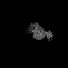 46691MC 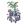 8zufC 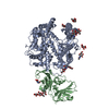 9c6oC M: map data used to model this data C: citing same article ( |
|---|
-Links
 PDBj
PDBj- Assembly
| Deposited unit | 
|
|---|---|
| 1 |
|
-Components
-Protein , 2 types, 2 molecules AB
| #1: Protein | Mass: 102102.375 Da / Num. of mol.: 1 Fragment: GenBank CAK6450646.1 chimeric sequence with Bat30 (Pteronotus davyi) residues 407-432, 598; includes N-terminal signal sequence and C-terminal tag for protein expression Source method: isolated from a genetically manipulated source Details: N-terminal tag: MGGEGLRASPRRRPLLPLQPRGCPRGDGCLRGGRGRAGFGFWRVTGGSRASANHVHAFFFFLQLLGNVLVIVLSHHFGKEFATMPMGSLQPLATLYLLGMLVASVLA C-terminal tag: ...Details: N-terminal tag: MGGEGLRASPRRRPLLPLQPRGCPRGDGCLRGGRGRAGFGFWRVTGGSRASANHVHAFFFFLQLLGNVLVIVLSHHFGKEFATMPMGSLQPLATLYLLGMLVASVLA C-terminal tag: PTLGPPYQPPVTGTGGGSWSHPQFEKGGGSGGGSGGSAWSHPQFEKGGGGSDYKDHDGDYKDHDIDYKDDDDKL Source: (gene. exp.) Pipistrellus nathusii (Nathusius's pipistrelle)Production host:  Homo sapiens (human) Homo sapiens (human) |
|---|---|
| #2: Protein | Mass: 34357.488 Da / Num. of mol.: 1 Fragment: GenBank WDE20340.1; includes N-terminal signal sequence and C-terminal tag for protein expression Source method: isolated from a genetically manipulated source Details: N-terminal tag: MGILPSPGMPALLSLVSLLSVLLMGCVAETGT C-terminal tag: LVPRGSSSGGSGLNDIFEAQKIEWHEGGSHHHHHHHH Source: (gene. exp.) Merbecovirus / Strain: PnNL2018B / Production host: Homo sapiens (human) |
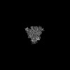
-Sugars , 4 types, 11 molecules 
| #3: Polysaccharide | Source method: isolated from a genetically manipulated source #4: Polysaccharide | Source method: isolated from a genetically manipulated source #5: Polysaccharide | Source method: isolated from a genetically manipulated source #6: Sugar | ChemComp-NAG /  Type: D-saccharide, beta linking / Mass: 221.208 Da / Num. of mol.: 4 / Source method: obtained synthetically / Formula: C8H15NO6 Type: D-saccharide, beta linking / Mass: 221.208 Da / Num. of mol.: 4 / Source method: obtained synthetically / Formula: C8H15NO6 |
|---|
-Non-polymers , 2 types, 145 molecules 

| #7: Chemical | ChemComp-ZN /  Mass: 65.409 Da / Num. of mol.: 1 / Source method: obtained synthetically / Formula: Zn Mass: 65.409 Da / Num. of mol.: 1 / Source method: obtained synthetically / Formula: Zn |
|---|---|
| #8: Water | ChemComp-HOH / Mass: 18.015 Da / Num. of mol.: 144 / Source method: isolated from a natural source / Formula: H2O |
-Details
| Has ligand of interest | N |
|---|---|
| Has protein modification | Y |
-Experimental details
-Experiment
| Experiment | Method: ELECTRON MICROSCOPY |
|---|---|
| EM experiment | Aggregation state: PARTICLE / 3D reconstruction method: single particle reconstruction |
- Sample preparation
| Component | Name: Merbecovirus PnNL2018B Spike glycoprotein RBD bound to the P. Nathusii ACE2 Type: COMPLEX / Entity ID: #1-#2 / Source: MULTIPLE SOURCES |
|---|---|
| Molecular weight | Experimental value: NO |
| Source (natural) | Organism: Merbecovirus |
| Source (recombinant) | Organism: Homo sapiens (human) |
| Buffer solution | pH: 8 |
| Specimen | Embedding applied: NO / Shadowing applied: NO / Staining applied: NO / Vitrification applied: YES |
| Vitrification | Cryogen name: ETHANE |
- Electron microscopy imaging
| Experimental equipment |  Model: Titan Krios / Image courtesy: FEI Company |
|---|---|
| Microscopy | Model: FEI TITAN KRIOS |
| Electron gun | Electron source:  FIELD EMISSION GUN / Accelerating voltage: 300 kV / Illumination mode: FLOOD BEAM FIELD EMISSION GUN / Accelerating voltage: 300 kV / Illumination mode: FLOOD BEAM |
| Electron lens | Mode: BRIGHT FIELD / Nominal defocus max: 3500 nm / Nominal defocus min: 200 nm |
| Image recording | Electron dose: 60 e/Å2 / Film or detector model: GATAN K3 (6k x 4k) |
- Processing
| EM software |
| |||||||||
|---|---|---|---|---|---|---|---|---|---|---|
| CTF correction | Type: PHASE FLIPPING AND AMPLITUDE CORRECTION | |||||||||
| 3D reconstruction | Resolution: 2.4 Å / Resolution method: FSC 0.143 CUT-OFF / Num. of particles: 1555546 / Symmetry type: POINT |
