 Movie
Movie Controller

[English] 日本語
 Yorodumi
- EMDB-60483: Cryo-EM structure of P.nat ACE2 mutant in complex with MOW15-22 RBD -
+ Open data

- Basic information
| Entry |  | |||||||||
|---|---|---|---|---|---|---|---|---|---|---|
| Title | Cryo-EM structure of P.nat ACE2 mutant in complex with MOW15-22 RBD | |||||||||
 Map data Map data | ||||||||||
 Sample Sample |
| |||||||||
 Keywords Keywords | ACE2 / RBD / VIRAL PROTEIN/HYDROLASE / VIRAL PROTEIN-HYDROLASE complex | |||||||||
| Biological species |  Pipistrellus nathusii (Nathusius's pipistrelle) / Pipistrellus nathusii (Nathusius's pipistrelle) /   Middle East respiratory syndrome-related coronavirus Middle East respiratory syndrome-related coronavirus | |||||||||
| Method | single particle reconstruction / cryo EM / Resolution: 3.31 Å | |||||||||
 Authors Authors | Tang J / Deng Z | |||||||||
| Funding support | 1 items
| |||||||||
 Citation Citation | Journal: Cell / Year: 2025 Title: Multiple independent acquisitions of ACE2 usage in MERS-related coronaviruses. Authors: Cheng-Bao Ma / Chen Liu / Young-Jun Park / Jingjing Tang / Jing Chen / Qing Xiong / Jimin Lee / Cameron Stewart / Daniel Asarnow / Jack Brown / M Alejandra Tortorici / Xiao Yang / Ye-Hui Sun ...Authors: Cheng-Bao Ma / Chen Liu / Young-Jun Park / Jingjing Tang / Jing Chen / Qing Xiong / Jimin Lee / Cameron Stewart / Daniel Asarnow / Jack Brown / M Alejandra Tortorici / Xiao Yang / Ye-Hui Sun / Yuan-Mei Chen / Xiao Yu / Jun-Yu Si / Peng Liu / Fei Tong / Mei-Ling Huang / Jing Li / Zheng-Li Shi / Zengqin Deng / David Veesler / Huan Yan /   Abstract: The angiotensin-converting enzyme 2 (ACE2) receptor is shared by various coronaviruses with distinct receptor-binding domain (RBD) architectures, yet our understanding of these convergent acquisition ...The angiotensin-converting enzyme 2 (ACE2) receptor is shared by various coronaviruses with distinct receptor-binding domain (RBD) architectures, yet our understanding of these convergent acquisition events remains elusive. Here, we report that two bat MERS-related coronaviruses (MERSr-CoVs) infecting Pipistrellus nathusii (P.nat)-MOW15-22 and PnNL2018B-use ACE2 as their receptor, with narrow ortholog specificity. Cryoelectron microscopy structures of the MOW15-22/PnNL2018B RBD-ACE2 complexes unveil an unexpected and entirely distinct binding mode, mapping >45 Å away from that of any other known ACE2-using coronaviruses. Functional profiling of ACE2 orthologs from 105 mammalian species led to the identification of host tropism determinants, including an ACE2 N432-glycosylation restricting viral recognition, and the design of a soluble P.nat ACE2 mutant with potent viral neutralizing activity. Our findings reveal convergent acquisition of ACE2 usage for merbecoviruses found in European bats, underscoring the extraordinary diversity of ACE2 recognition modes among coronaviruses and the promiscuity of this receptor. | |||||||||
| History |
|
- Structure visualization
| Supplemental images |
|---|

- Downloads & links
-EMDB archive
| Map data | emd_60483.map.gz | 28.8 MB |  EMDB map data format EMDB map data format | |
|---|---|---|---|---|
| Header (meta data) | emd-60483-v30.xmlemd-60483.xml | 18.9 KB 18.9 KB | Display Display | EMDB header |
| Images |  emd_60483.png emd_60483.png | 89 KB | ||
| Filedesc metadata | emd-60483.cif.gz | 6.8 KB | ||
| Others | emd_60483_half_map_1.map.gzemd_60483_half_map_2.map.gz | 28.3 MB 28.3 MB | ||
| Archive directory |  http://ftp.pdbj.org/pub/emdb/structures/EMD-60483ftp://ftp.pdbj.org/pub/emdb/structures/EMD-60483 http://ftp.pdbj.org/pub/emdb/structures/EMD-60483ftp://ftp.pdbj.org/pub/emdb/structures/EMD-60483 | HTTPS FTP |
-Related structure data
| Related structure data | 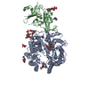 8zufMC 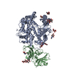 9c6oC 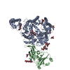 9dakC M: atomic model generated by this map C: citing same article ( |
|---|
-Links
| EMDB pages | EMDB (EBI/PDBe) / EMDataResource |
|---|
-Map
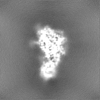
| File | Download / File: emd_60483.map.gz / Format: CCP4 / Size: 30.5 MB / Type: IMAGE STORED AS FLOATING POINT NUMBER (4 BYTES) | ||||||||||||||||||||||||||||||||||||
|---|---|---|---|---|---|---|---|---|---|---|---|---|---|---|---|---|---|---|---|---|---|---|---|---|---|---|---|---|---|---|---|---|---|---|---|---|---|
| Projections & slices | Image control
Images are generated by Spider. | ||||||||||||||||||||||||||||||||||||
| Voxel size | X=Y=Z: 0.95 Å | ||||||||||||||||||||||||||||||||||||
| Density |
| ||||||||||||||||||||||||||||||||||||
| Symmetry | Space group: 1 | ||||||||||||||||||||||||||||||||||||
| Details | EMDB XML:
|
 Z (Sec.)
 Y (Row.)
 X (Col.)


-Supplemental data
-Half map: #2
| File | emd_60483_half_map_1.map | ||||||||||||
|---|---|---|---|---|---|---|---|---|---|---|---|---|---|
| Projections & Slices |
| ||||||||||||
| Density Histograms |


-Half map: #1
| File | emd_60483_half_map_2.map | ||||||||||||
|---|---|---|---|---|---|---|---|---|---|---|---|---|---|
| Projections & Slices |
| ||||||||||||
| Density Histograms |


- Sample components
-Entire : P.nat ACE2-MOW15-22 RBD
| Entire | Name: P.nat ACE2-MOW15-22 RBD |
|---|---|
| Components |
|
-Supramolecule #1: P.nat ACE2-MOW15-22 RBD
| Supramolecule | Name: P.nat ACE2-MOW15-22 RBD / type: complex / ID: 1 / Parent: 0 / Macromolecule list: #1-#2 |
|---|---|
| Source (natural) | Organism: Pipistrellus nathusii (Nathusius's pipistrelle) |
-Macromolecule #1: Angiotensin-converting enzyme
| Macromolecule | Name: Angiotensin-converting enzyme / type: protein_or_peptide / ID: 1 / Number of copies: 1 / Enantiomer: LEVO |
|---|---|
| Source (natural) | Organism: Pipistrellus nathusii (Nathusius's pipistrelle) |
| Molecular weight | Theoretical: 90.767672 KDa |
| Recombinant expression | Organism:  Homo sapiens (human) Homo sapiens (human) |
| Sequence | String: EEKAREFLDK FNSEAENWSH ESALASWDYN TNINDKNAQK MNEADSKWSA FYKEHSKLAQ GFPLQEIQNS TIKLQLQILQ QNGSSVLTA EKSKRLSTIL TTMSTIYSTG KVCNPNNPQQ CFTLSGLEDI MEKSKDYHQR LWIWEGWRSE VGKQLRPLYE E YVALKNEM ...String: EEKAREFLDK FNSEAENWSH ESALASWDYN TNINDKNAQK MNEADSKWSA FYKEHSKLAQ GFPLQEIQNS TIKLQLQILQ QNGSSVLTA EKSKRLSTIL TTMSTIYSTG KVCNPNNPQQ CFTLSGLEDI MEKSKDYHQR LWIWEGWRSE VGKQLRPLYE E YVALKNEM ARGNNYKDYG DYWRGDYETE GGDGYNYSRN HLIEDVDRIF LEIKPLYEQL HAYVRAKLMN AYPSRISPTG CL PAHLLGD MWGRFWTNLY NLTVPFEKKQ NIDVTDTMKK QSWDAEKIFK EAEKFYLSVG LHNMTPEFWN NSMLTEPSDG RQV VCHPTA WDLGKNDFRI KMCTKVTMDD FLTAHHEMGH IQYDMAYAKQ PYLLRNGANE GFHEAVGEIM SLSAATPKHL KDLG LLAQN YPEDYETEIN FLLKQALNIV GTLPFTYMLE KWRWMVFEGK IPKEQWMEKW WEMKREIVGV VEPLPHDETY CDPAS LFHV ANDYSFIRYF TRTILEFQFQ EALCQIANHT GPLHKCDISN STEAGKQLKN MLELGKSKPW TFALEQIART KEMDAK PLL NYFKPLFSWL KELNGNSVGW SADWSPYSEQ SIKVRISLKS ALGEKAYEWN DNEMYLFRSS VAYAMRVYFL KVKNETI PF RAEDVWVSDE KIRVSFKFFV TSPTNVSDII PRSEVEDAIR MSRGRINDAF RLDDKTLEFL GIQPTLGPPY QPPVTIWL I VFGVVMGMVV IGIGVLIFTG IRDRKKKNQA ENEENPYSSV NLSKGENNPG FQSGDDVQTS F |
-Macromolecule #2: MOW15-22 RBD
| Macromolecule | Name: MOW15-22 RBD / type: protein_or_peptide / ID: 2 / Number of copies: 1 / Enantiomer: LEVO |
|---|---|
| Source (natural) | Organism: Middle East respiratory syndrome-related coronavirus |
| Molecular weight | Theoretical: 23.646434 KDa |
| Recombinant expression | Organism: Homo sapiens (human) |
| Sequence | String: ECDFTKLFIG QVPQPYEFGR LVFTNCNYNF TKLLSYFQVN TFQCQKVTPE SIATGCYSSL TVDWFAYRVE DKSDLLPGSS SDLQRFNYK PTYSNPTCLI SAYTNLVPLG GVNPTNYTTL TNCYGCVDKD PANPWGDQIC IPEFVTEVEP GFRPKPSCAR V GLEGHISG ...String: ECDFTKLFIG QVPQPYEFGR LVFTNCNYNF TKLLSYFQVN TFQCQKVTPE SIATGCYSSL TVDWFAYRVE DKSDLLPGSS SDLQRFNYK PTYSNPTCLI SAYTNLVPLG GVNPTNYTTL TNCYGCVDKD PANPWGDQIC IPEFVTEVEP GFRPKPSCAR V GLEGHISG NDTYSAIVTN GELDSTGDPI WRKGVALTKQ PIDSSRADLA FFVSV |
-Macromolecule #5: 2-acetamido-2-deoxy-beta-D-glucopyranose
| Macromolecule | Name: 2-acetamido-2-deoxy-beta-D-glucopyranose / type: ligand / ID: 5 / Number of copies: 4 / Formula: NAG |
|---|---|
| Molecular weight | Theoretical: 221.208 Da |
| Chemical component information |  ChemComp-NAG: |
-Macromolecule #6: ZINC ION
| Macromolecule | Name: ZINC ION / type: ligand / ID: 6 / Number of copies: 1 / Formula: ZN |
|---|---|
| Molecular weight | Theoretical: 65.409 Da |
-Experimental details
-Structure determination
| Method | cryo EM |
|---|---|
 Processing Processing | single particle reconstruction |
| Aggregation state | particle |
-Sample preparation
| Buffer | pH: 8 |
|---|---|
| Vitrification | Cryogen name: ETHANE |
- Electron microscopy
| Microscope | JEOL CRYO ARM 300 |
|---|---|
| Image recording | Film or detector model: GATAN K3 (6k x 4k) / Average electron dose: 40.0 e/Å2 |
| Electron beam | Acceleration voltage: 300 kV / Electron source:  FIELD EMISSION GUN FIELD EMISSION GUN |
| Electron optics | Illumination mode: FLOOD BEAM / Imaging mode: BRIGHT FIELD / Nominal defocus max: 2.5 µm / Nominal defocus min: 0.5 µm |
