Movie
Movie Controller

[English] 日本語
 Yorodumi
- PDB-9cth: Preliminary map of the Prothrombin-prothrombinase complex on nano... -
+ Open data

- Basic information
| Entry | Database: PDB / ID: 9cth | ||||||||||||||||||
|---|---|---|---|---|---|---|---|---|---|---|---|---|---|---|---|---|---|---|---|
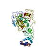
| Title | Preliminary map of the Prothrombin-prothrombinase complex on nano discs | ||||||||||||||||||
 Components Components |
| ||||||||||||||||||
 Keywords Keywords | BLOOD CLOTTING / Coagulation / Prothrombin / Prothrombinase / nanodisc / complex | ||||||||||||||||||
| Function / homology |  Function and homology information Function and homology informationresponse to vitamin K / coagulation factor Xa / platelet alpha granule / Cargo concentration in the ER / Defective factor IX causes thrombophilia / Defective cofactor function of FVIIIa variant / Defective F9 variant does not activate FX / COPII-coated ER to Golgi transport vesicle / : / COPII-mediated vesicle transport ...response to vitamin K / coagulation factor Xa / platelet alpha granule / Cargo concentration in the ER / Defective factor IX causes thrombophilia / Defective cofactor function of FVIIIa variant / Defective F9 variant does not activate FX / COPII-coated ER to Golgi transport vesicle / : / COPII-mediated vesicle transport / : / blood circulation / thrombospondin receptor activity / thrombin / thrombin-activated receptor signaling pathway / Defective factor XII causes hereditary angioedema / negative regulation of astrocyte differentiation / regulation of blood coagulation / neutrophil-mediated killing of gram-negative bacterium / positive regulation of phospholipase C-activating G protein-coupled receptor signaling pathway / Defective F8 cleavage by thrombin / Platelet Aggregation (Plug Formation) / ligand-gated ion channel signaling pathway / positive regulation of collagen biosynthetic process / negative regulation of platelet activation / negative regulation of blood coagulation / negative regulation of fibrinolysis / blood coagulation, fibrin clot formation / positive regulation of blood coagulation / positive regulation of TOR signaling / Transport of gamma-carboxylated protein precursors from the endoplasmic reticulum to the Golgi apparatus / : / Gamma-carboxylation of protein precursors / Removal of aminoterminal propeptides from gamma-carboxylated proteins / regulation of cytosolic calcium ion concentration / fibrinolysis / : / negative regulation of proteolysis / endoplasmic reticulum-Golgi intermediate compartment membrane / negative regulation of cytokine production involved in inflammatory response / platelet alpha granule lumen / Regulation of Complement cascade / acute-phase response / Cell surface interactions at the vascular wall / positive regulation of release of sequestered calcium ion into cytosol / Peptide ligand-binding receptors / growth factor activity / positive regulation of receptor signaling pathway via JAK-STAT / Post-translational protein phosphorylation / lipopolysaccharide binding / platelet activation / phospholipid binding / response to wounding / positive regulation of protein localization to nucleus / Golgi lumen / Regulation of Insulin-like Growth Factor (IGF) transport and uptake by Insulin-like Growth Factor Binding Proteins (IGFBPs) / positive regulation of reactive oxygen species metabolic process / blood coagulation / positive regulation of insulin secretion / Platelet degranulation / regulation of cell shape / antimicrobial humoral immune response mediated by antimicrobial peptide / heparin binding / extracellular vesicle / Thrombin signalling through proteinase activated receptors (PARs) / positive regulation of cell growth / blood microparticle / G alpha (q) signalling events / cell surface receptor signaling pathway / positive regulation of phosphatidylinositol 3-kinase/protein kinase B signal transduction / positive regulation of cell migration / endoplasmic reticulum lumen / receptor ligand activity / copper ion binding / signaling receptor binding / serine-type endopeptidase activity / external side of plasma membrane / calcium ion binding / positive regulation of cell population proliferation / proteolysis / : / extracellular exosome / extracellular region / membrane / plasma membrane Similarity search - Function | ||||||||||||||||||
| Biological species |  Homo sapiens (human) Homo sapiens (human) | ||||||||||||||||||
| Method | ELECTRON MICROSCOPY / single particle reconstruction / cryo EM / Resolution: 6.47 Å | ||||||||||||||||||
 Authors Authors | Stojanovski, B.M. / Mohammed, B.M. / Di Cera, E. | ||||||||||||||||||
| Funding support |  United States, 5items United States, 5items
| ||||||||||||||||||
 Citation Citation | Journal: Subcell Biochem / Year: 2024 Title: The Prothrombin-Prothrombinase Interaction. Authors: Bosko M Stojanovski / Bassem M Mohammed / Enrico Di Cera / Abstract: The hemostatic response to vascular injury entails a sequence of proteolytic events where several inactive zymogens of the trypsin family are converted to active proteases. The cascade starts with ...The hemostatic response to vascular injury entails a sequence of proteolytic events where several inactive zymogens of the trypsin family are converted to active proteases. The cascade starts with exposure of tissue factor from the damaged endothelium and culminates with conversion of prothrombin to thrombin in a reaction catalyzed by the prothrombinase complex composed of the enzyme factor Xa, cofactor Va, Ca, and phospholipids. This cofactor-dependent activation is paradigmatic of analogous reactions of the blood coagulation and complement cascades, which makes elucidation of its molecular mechanism of broad significance to the large class of trypsin-like zymogens to which prothrombin belongs. Because of its relevance as the most important reaction in the physiological response to vascular injury, as well as the main trigger of pathological thrombotic complications, the mechanism of prothrombin activation has been studied extensively. However, a molecular interpretation of this mechanism has become available only recently from important developments in structural biology. Here we review current knowledge on the prothrombin-prothrombinase interaction and outline future directions for the study of this key reaction of the coagulation cascade. | ||||||||||||||||||
| History |
|
- Structure visualization
| Structure viewer | Molecule: MolmilJmol/JSmol |
|---|
- Downloads & links
-Download
| PDBx/mmCIF format | 9cth.cif.gz | 418.5 KB | Display | PDBx/mmCIF format |
|---|---|---|---|---|
| PDB format | pdb9cth.ent.gz | Display | PDB format | |
| PDBx/mmJSON format | 9cth.json.gz | Tree view | PDBx/mmJSON format | |
| Others |  Other downloads Other downloads |
-Validation report
| Arichive directory | https://data.pdbj.org/pub/pdb/validation_reports/ct/9cthftp://data.pdbj.org/pub/pdb/validation_reports/ct/9cth | HTTPS FTP |
|---|
-Related structure data
| Related structure data | 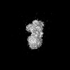 42405MC M: map data used to model this data C: citing same article ( |
|---|---|
| Similar structure data |

-Links
 PDBj
PDBj


- Assembly
| Deposited unit | 
|
|---|---|
| 1 |
|
-Components
-Activated Factor V (FVa) ... , 2 types, 2 molecules AE
| #1: Protein | Mass: 81274.391 Da / Num. of mol.: 1 / Fragment: Domains A1 and A2 (UNP residues 29-737) / Source method: isolated from a natural source / Source: (natural) Homo sapiens (human) / Tissue: Blood / References: UniProt: P12259 |
|---|---|
| #2: Protein | Mass: 75283.008 Da / Num. of mol.: 1 / Fragment: Domains C1, C2, and A3 (UNP residues 1574-2224) / Source method: isolated from a natural source / Source: (natural) Homo sapiens (human) / Tissue: Blood / References: UniProt: P12259 |
-Activated Factor X ... , 2 types, 2 molecules BC
| #3: Protein | Mass: 16186.952 Da / Num. of mol.: 1 Source method: isolated from a genetically manipulated source Source: (gene. exp.) Homo sapiens (human) / Gene: F10 / Cell (production host): fibroblasts / Cell line (production host): BHK / Production host:  Mesocricetus auratus (golden hamster) / References: UniProt: P00742 Mesocricetus auratus (golden hamster) / References: UniProt: P00742 |
|---|---|
| #4: Protein | Mass: 26588.297 Da / Num. of mol.: 1 / Mutation: S195A Source method: isolated from a genetically manipulated source Source: (gene. exp.) Homo sapiens (human) / Gene: F10 / Cell (production host): fibroblasts / Cell line (production host): BHK / Production host: Mesocricetus auratus (golden hamster) / References: UniProt: P00742 |
-Protein / Sugars , 2 types, 7 molecules D
| #5: Protein | Mass: 65370.113 Da / Num. of mol.: 1 / Fragment: UNP residues 44-622 / Mutation: S525A Source method: isolated from a genetically manipulated source Source: (gene. exp.) Homo sapiens (human) / Gene: F2 / Cell (production host): fibroblasts / Cell line (production host): BHK / Production host: Mesocricetus auratus (golden hamster) / References: UniProt: P00734, thrombin |
|---|---|
| #6: Sugar | ChemComp-NAG /  Type: D-saccharide, beta linking / Mass: 221.208 Da / Num. of mol.: 6 / Source method: obtained synthetically / Formula: C8H15NO6 / Feature type: SUBJECT OF INVESTIGATION Type: D-saccharide, beta linking / Mass: 221.208 Da / Num. of mol.: 6 / Source method: obtained synthetically / Formula: C8H15NO6 / Feature type: SUBJECT OF INVESTIGATION |
-Details
| Has ligand of interest | Y |
|---|---|
| Has protein modification | Y |
-Experimental details
-Experiment
| Experiment | Method: ELECTRON MICROSCOPY |
|---|---|
| EM experiment | Aggregation state: PARTICLE / 3D reconstruction method: single particle reconstruction |
- Sample preparation
| Component | Name: Prothrombinase:Prothrombin ternary complex on lipid nanodiscs Type: COMPLEX Details: The complex is made of coagulation factor Va (derived from human plasma), active site mutated (S195A) coagulation factor Xa (Recombinantly expressed in BHK cells) and active site mutated ...Details: The complex is made of coagulation factor Va (derived from human plasma), active site mutated (S195A) coagulation factor Xa (Recombinantly expressed in BHK cells) and active site mutated (S525A) Prothrombin (Recombinantly expressed in BHK cells). The nanodisc component of the complex is made of the scaffold protein MSP1E3D1 (Recombinantly expressed in bacteria) and the phospholipid component was Porcine Brain phosphatidylserine. Entity ID: #3-#5 / Source: RECOMBINANT |
|---|---|
| Molecular weight | Experimental value: NO |
| Source (natural) | Organism: Homo sapiens (human) |
| Source (recombinant) | Organism: Mesocricetus auratus (golden hamster) |
| Buffer solution | pH: 7.4 / Details: 20 mM HEPES, 150 mM NaCl, and 5 mM CaCl2 |
| Specimen | Conc.: 0.01 mg/ml / Embedding applied: NO / Shadowing applied: NO / Staining applied: NO / Vitrification applied: YES Details: Prothrombin:FVa:FXa were mixed in 2:1:2 ratio. 0.01 mg/mL total protein was used for freezing. |
| Specimen support | Grid material: COPPER / Grid mesh size: 300 divisions/in. / Grid type: Quantifoil R2/2 |
| Vitrification | Instrument: FEI VITROBOT MARK IV / Cryogen name: ETHANE / Humidity: 100 % |
- Electron microscopy imaging
| Microscopy | Model: TFS GLACIOS |
|---|---|
| Electron gun | Electron source:  FIELD EMISSION GUN / Accelerating voltage: 200 kV / Illumination mode: FLOOD BEAM FIELD EMISSION GUN / Accelerating voltage: 200 kV / Illumination mode: FLOOD BEAM |
| Electron lens | Mode: BRIGHT FIELD / Nominal defocus max: 2400 nm / Nominal defocus min: 800 nm / Cs: 2.7 mm / C2 aperture diameter: 50 µm |
| Specimen holder | Cryogen: NITROGEN |
| Image recording | Electron dose: 51.28 e/Å2 / Film or detector model: FEI FALCON IV (4k x 4k) / Num. of real images: 2390 |
- Processing
| EM software |
| ||||||||||||||||||||||||||||||||||||||||||
|---|---|---|---|---|---|---|---|---|---|---|---|---|---|---|---|---|---|---|---|---|---|---|---|---|---|---|---|---|---|---|---|---|---|---|---|---|---|---|---|---|---|---|---|
| CTF correction | Type: PHASE FLIPPING AND AMPLITUDE CORRECTION | ||||||||||||||||||||||||||||||||||||||||||
| Symmetry | Point symmetry: C1 (asymmetric) | ||||||||||||||||||||||||||||||||||||||||||
| 3D reconstruction | Resolution: 6.47 Å / Resolution method: FSC 0.143 CUT-OFF / Num. of particles: 4988 / Algorithm: BACK PROJECTION / Symmetry type: POINT | ||||||||||||||||||||||||||||||||||||||||||
| Atomic model building | Protocol: FLEXIBLE FIT / Space: REAL | ||||||||||||||||||||||||||||||||||||||||||
| Atomic model building |
| ||||||||||||||||||||||||||||||||||||||||||
| Refine LS restraints |
|