Movie
Movie Controller

[English] 日本語
 Yorodumi
- PDB-9crd: Cryo-EM structure of SARS-CoV-2 Spike Proteins on intact virions:... -
+ Open data

- Basic information
| Entry | Database: PDB / ID: 9crd | ||||||||||||
|---|---|---|---|---|---|---|---|---|---|---|---|---|---|
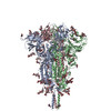
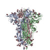
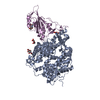
| Title | Cryo-EM structure of SARS-CoV-2 Spike Proteins on intact virions: B.1 variant 1 open RBD | ||||||||||||
 Components Components | Spike glycoprotein | ||||||||||||
 Keywords Keywords | VIRAL PROTEIN / SARS-CoV-2 variant / B.1 strain | ||||||||||||
| Function / homology |  Function and homology information Function and homology informationsymbiont-mediated disruption of host tissue / Maturation of spike protein / Translation of Structural Proteins / Virion Assembly and Release / host cell surface / host extracellular region / symbiont-mediated-mediated suppression of host tetherin activity / Induction of Cell-Cell Fusion / structural constituent of virion / positive regulation of viral entry into host cell ...symbiont-mediated disruption of host tissue / Maturation of spike protein / Translation of Structural Proteins / Virion Assembly and Release / host cell surface / host extracellular region / symbiont-mediated-mediated suppression of host tetherin activity / Induction of Cell-Cell Fusion / structural constituent of virion / positive regulation of viral entry into host cell / membrane fusion / host cell endoplasmic reticulum-Golgi intermediate compartment membrane / Attachment and Entry / entry receptor-mediated virion attachment to host cell / receptor-mediated virion attachment to host cell / host cell surface receptor binding / symbiont-mediated suppression of host innate immune response / endocytosis involved in viral entry into host cell / receptor ligand activity / fusion of virus membrane with host plasma membrane / fusion of virus membrane with host endosome membrane / viral envelope / symbiont entry into host cell / virion attachment to host cell / host cell plasma membrane / SARS-CoV-2 activates/modulates innate and adaptive immune responses / virion membrane / membrane / identical protein binding / plasma membrane Similarity search - Function | ||||||||||||
| Biological species |   Severe acute respiratory syndrome coronavirus 2 Severe acute respiratory syndrome coronavirus 2 | ||||||||||||
| Method | ELECTRON MICROSCOPY / single particle reconstruction / cryo EM / Resolution: 3.5 Å | ||||||||||||
 Authors Authors | Ke, Z. / Croll, T.I. / Briggs, J.A.G. | ||||||||||||
| Funding support |  United Kingdom, European Union, United Kingdom, European Union,  Germany, 3items Germany, 3items
| ||||||||||||
 Citation Citation | Journal: EMBO J / Year: 2024 Title: Virion morphology and on-virus spike protein structures of diverse SARS-CoV-2 variants. Authors: Zunlong Ke / Thomas P Peacock / Jonathan C Brown / Carol M Sheppard / Tristan I Croll / Abhay Kotecha / Daniel H Goldhill / Wendy S Barclay / John A G Briggs /   Abstract: The evolution of SARS-CoV-2 variants with increased fitness has been accompanied by structural changes in the spike (S) proteins, which are the major target for the adaptive immune response. Single- ...The evolution of SARS-CoV-2 variants with increased fitness has been accompanied by structural changes in the spike (S) proteins, which are the major target for the adaptive immune response. Single-particle cryo-EM analysis of soluble S protein from SARS-CoV-2 variants has revealed this structural adaptation at high resolution. The analysis of S trimers in situ on intact virions has the potential to provide more functionally relevant insights into S structure and virion morphology. Here, we characterized B.1, Alpha, Beta, Gamma, Delta, Kappa, and Mu variants by cryo-electron microscopy and tomography, assessing S cleavage, virion morphology, S incorporation, "in-situ" high-resolution S structures, and the range of S conformational states. We found no evidence for adaptive changes in virion morphology, but describe multiple different positions in the S protein where amino acid changes alter local protein structure. Taken together, our data are consistent with a model where amino acid changes at multiple positions from the top to the base of the spike cause structural changes that can modulate the conformational dynamics of the S protein. | ||||||||||||
| History |
|
- Structure visualization
| Structure viewer | Molecule: MolmilJmol/JSmol |
|---|
- Downloads & links
-Download
| PDBx/mmCIF format | 9crd.cif.gz | 634.8 KB | Display | PDBx/mmCIF format |
|---|---|---|---|---|
| PDB format | pdb9crd.ent.gz | 517.5 KB | Display | PDB format |
| PDBx/mmJSON format | 9crd.json.gz | Tree view | PDBx/mmJSON format | |
| Others |  Other downloads Other downloads |
-Validation report
| Arichive directory | https://data.pdbj.org/pub/pdb/validation_reports/cr/9crdftp://data.pdbj.org/pub/pdb/validation_reports/cr/9crd | HTTPS FTP |
|---|
-Related structure data
| Related structure data |  45864MC  9crcC  9creC  9crfC  9crgC  9crhC  9criC C: citing same article ( M: map data used to model this data |
|---|---|
| Similar structure data |


-Links
 PDBj
PDBj


- Assembly
| Deposited unit | 
|
|---|---|
| 1 |
|
-Components
| #1: Protein | Mass: 141253.422 Da / Num. of mol.: 3 / Source method: isolated from a natural source Source: (natural) Severe acute respiratory syndrome coronavirus 2References: UniProt: P0DTC2 #2: Polysaccharide | 2-acetamido-2-deoxy-beta-D-glucopyranose-(1-4)-2-acetamido-2-deoxy-beta-D-glucopyranose Source method: isolated from a genetically manipulated source #3: Polysaccharide | Source method: isolated from a genetically manipulated source #4: Sugar | ChemComp-NAG /   Type: D-saccharide, beta linking / Mass: 221.208 Da / Num. of mol.: 19 / Source method: obtained synthetically / Formula: C8H15NO6 Type: D-saccharide, beta linking / Mass: 221.208 Da / Num. of mol.: 19 / Source method: obtained synthetically / Formula: C8H15NO6Has ligand of interest | N | Has protein modification | Y | |
|---|
-Experimental details
-Experiment
| Experiment | Method: ELECTRON MICROSCOPY |
|---|---|
| EM experiment | Aggregation state: PARTICLE / 3D reconstruction method: single particle reconstruction |
- Sample preparation
| Component | Name: Severe acute respiratory syndrome coronavirus 2 / Type: VIRUS / Entity ID: #1 / Source: NATURAL |
|---|---|
| Source (natural) | Organism: Severe acute respiratory syndrome coronavirus 2 / Strain: B.1 |
| Source (recombinant) | Organism:  Homo sapiens (human) Homo sapiens (human) |
| Details of virus | Empty: NO / Enveloped: YES / Isolate: STRAIN / Type: VIRION |
| Natural host | Organism: Homo sapiens |
| Buffer solution | pH: 7 |
| Specimen | Embedding applied: NO / Shadowing applied: NO / Staining applied: NO / Vitrification applied: YES |
| Vitrification | Cryogen name: ETHANE |
- Electron microscopy imaging
| Experimental equipment |  Model: Titan Krios / Image courtesy: FEI Company |
|---|---|
| Microscopy | Model: FEI TITAN KRIOS |
| Electron gun | Electron source:  FIELD EMISSION GUN / Accelerating voltage: 300 kV / Illumination mode: FLOOD BEAM FIELD EMISSION GUN / Accelerating voltage: 300 kV / Illumination mode: FLOOD BEAM |
| Electron lens | Mode: BRIGHT FIELD / Nominal defocus max: 3000 nm / Nominal defocus min: 1000 nm / Calibrated defocus min: 1000 nm / Calibrated defocus max: 3000 nm / Cs: 2.7 mm |
| Specimen holder | Cryogen: NITROGEN / Specimen holder model: FEI TITAN KRIOS AUTOGRID HOLDER |
| Image recording | Electron dose: 50 e/Å2 / Film or detector model: GATAN K3 BIOQUANTUM (6k x 4k) / Num. of real images: 5598 |
- Processing
| EM software |
| ||||||||||||||||||||
|---|---|---|---|---|---|---|---|---|---|---|---|---|---|---|---|---|---|---|---|---|---|
| CTF correction | Type: PHASE FLIPPING AND AMPLITUDE CORRECTION | ||||||||||||||||||||
| Particle selection | Num. of particles selected: 908151 | ||||||||||||||||||||
| Symmetry | Point symmetry: C1 (asymmetric) | ||||||||||||||||||||
| 3D reconstruction | Resolution: 3.5 Å / Resolution method: FSC 0.143 CUT-OFF / Num. of particles: 37829 / Algorithm: BACK PROJECTION / Symmetry type: POINT | ||||||||||||||||||||
| EM volume selection | Num. of tomograms: 42 / Num. of volumes extracted: 400000 | ||||||||||||||||||||
| Atomic model building | Protocol: FLEXIBLE FIT | ||||||||||||||||||||
| Atomic model building | 3D fitting-ID: 1 / Source name: PDB / Type: experimental model
|