 Movie
Movie Controller

[English] 日本語
 Yorodumi
- PDB-9cc0: Human Mitochondrial LONP1 Degrading Casein, ATP-bound closed form -
+ Open data

- Basic information
| Entry | Database: PDB / ID: 9cc0 | |||||||||
|---|---|---|---|---|---|---|---|---|---|---|
| Title | Human Mitochondrial LONP1 Degrading Casein, ATP-bound closed form | |||||||||
 Components Components |
| |||||||||
 Keywords Keywords | HYDROLASE / ATPase / protease | |||||||||
| Function / homology |  Function and homology information Function and homology informationoxidation-dependent protein catabolic process / PH domain binding / mitochondrial protein catabolic process / G-quadruplex DNA binding / endopeptidase La / mitochondrial DNA metabolic process / mitochondrial genome maintenance / ATP-dependent peptidase activity / protein quality control for misfolded or incompletely synthesized proteins / mitochondrial nucleoid ...oxidation-dependent protein catabolic process / PH domain binding / mitochondrial protein catabolic process / G-quadruplex DNA binding / endopeptidase La / mitochondrial DNA metabolic process / mitochondrial genome maintenance / ATP-dependent peptidase activity / protein quality control for misfolded or incompletely synthesized proteins / mitochondrial nucleoid / regulation of peptidyl-tyrosine phosphorylation / insulin receptor substrate binding / chaperone-mediated protein complex assembly / DNA polymerase binding / negative regulation of insulin receptor signaling pathway / mitochondrion organization / proteolysis involved in protein catabolic process / Mitochondrial protein degradation / protein catabolic process / ADP binding / single-stranded DNA binding / cellular response to oxidative stress / sequence-specific DNA binding / single-stranded RNA binding / response to hypoxia / mitochondrial matrix / serine-type endopeptidase activity / ATP hydrolysis activity / mitochondrion / nucleoplasm / ATP binding / identical protein binding / membrane / cytosol Similarity search - Function | |||||||||
| Biological species |  Homo sapiens (human) Homo sapiens (human) | |||||||||
| Method | ELECTRON MICROSCOPY / single particle reconstruction / cryo EM / Resolution: 3.31 Å | |||||||||
 Authors Authors | Mindrebo, J.T. / Lander, G.C. | |||||||||
| Funding support |  United States, 2items United States, 2items
| |||||||||
 Citation Citation | Journal: bioRxiv / Year: 2024 Title: Structural and mechanistic studies on human LONP1 redefine the hand-over-hand translocation mechanism. Authors: Jeffrey T Mindrebo / Gabriel C Lander / Abstract: AAA+ enzymes use energy from ATP hydrolysis to remodel diverse cellular targets. Structures of substrate-bound AAA+ complexes suggest that these enzymes employ a conserved hand-over-hand mechanism to ...AAA+ enzymes use energy from ATP hydrolysis to remodel diverse cellular targets. Structures of substrate-bound AAA+ complexes suggest that these enzymes employ a conserved hand-over-hand mechanism to thread substrates through their central pore. However, the fundamental aspects of the mechanisms governing motor function and substrate processing within specific AAA+ families remain unresolved. We used cryo-electron microscopy to structurally interrogate reaction intermediates from in vitro biochemical assays to inform the underlying regulatory mechanisms of the human mitochondrial AAA+ protease, LONP1. Our results demonstrate that substrate binding allosterically regulates proteolytic activity, and that LONP1 can adopt a configuration conducive to substrate translocation even when the ATPases are bound to ADP. These results challenge the conventional understanding of the hand-over-hand translocation mechanism, giving rise to an alternative model that aligns more closely with biochemical and biophysical data on related enzymes like ClpX, ClpA, the 26S proteasome, and Lon protease. | |||||||||
| History |
|
- Structure visualization
| Structure viewer | Molecule: MolmilJmol/JSmol |
|---|
- Downloads & links
-Download
| PDBx/mmCIF format | 9cc0.cif.gz | 576.7 KB | Display | PDBx/mmCIF format |
|---|---|---|---|---|
| PDB format | pdb9cc0.ent.gz | 448.2 KB | Display | PDB format |
| PDBx/mmJSON format | 9cc0.json.gz | Tree view | PDBx/mmJSON format | |
| Others |  Other downloads Other downloads |
-Validation report
| Summary document | 9cc0_validation.pdf.gz | 1.9 MB | Display | wwPDB validaton report |
|---|---|---|---|---|
| Full document | 9cc0_full_validation.pdf.gz | 1.9 MB | Display | |
| Data in XML | 9cc0_validation.xml.gz | 91.7 KB | Display | |
| Data in CIF | 9cc0_validation.cif.gz | 136 KB | Display | |
| Arichive directory | https://data.pdbj.org/pub/pdb/validation_reports/cc/9cc0ftp://data.pdbj.org/pub/pdb/validation_reports/cc/9cc0 | HTTPS FTP |
-Related structure data
| Related structure data | 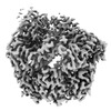 45430MC 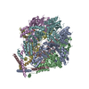 9cc3C M: map data used to model this data C: citing same article ( |
|---|---|
| Similar structure data |


-Links
 PDBj
PDBj
- Assembly
| Deposited unit | 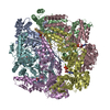
|
|---|---|
| 1 |
|
-Components
| #1: Protein | Mass: 97468.156 Da / Num. of mol.: 6 Source method: isolated from a genetically manipulated source Source: (gene. exp.) Homo sapiens (human) / Gene: LONP1, PRSS15 / Plasmid: pET-20aProduction host:  References: UniProt: P36776, endopeptidase La #2: Protein/peptide | | Mass: 1039.273 Da / Num. of mol.: 1 / Source method: isolated from a natural source / Source: (natural) #3: Chemical | ChemComp-ATP /   Mass: 507.181 Da / Num. of mol.: 4 / Source method: obtained synthetically / Formula: C10H16N5O13P3 / Feature type: SUBJECT OF INVESTIGATION / Comment: ATP, energy-carrying molecule*YM Mass: 507.181 Da / Num. of mol.: 4 / Source method: obtained synthetically / Formula: C10H16N5O13P3 / Feature type: SUBJECT OF INVESTIGATION / Comment: ATP, energy-carrying molecule*YM#4: Chemical | ChemComp-MG /   Mass: 24.305 Da / Num. of mol.: 4 / Source method: obtained synthetically / Formula: Mg Mass: 24.305 Da / Num. of mol.: 4 / Source method: obtained synthetically / Formula: Mg#5: Chemical |   Mass: 427.201 Da / Num. of mol.: 2 / Source method: obtained synthetically / Formula: C10H15N5O10P2 / Feature type: SUBJECT OF INVESTIGATION / Comment: ADP, energy-carrying molecule*YM Mass: 427.201 Da / Num. of mol.: 2 / Source method: obtained synthetically / Formula: C10H15N5O10P2 / Feature type: SUBJECT OF INVESTIGATION / Comment: ADP, energy-carrying molecule*YMHas ligand of interest | Y | Sequence details | Actual Crystalized sequence of Chain G is ...Actual Crystalized sequence of Chain G is MKVLILACLV | |
|---|
-Experimental details
-Experiment
| Experiment | Method: ELECTRON MICROSCOPY |
|---|---|
| EM experiment | Aggregation state: PARTICLE / 3D reconstruction method: single particle reconstruction |
- Sample preparation
| Component | Name: LONP1 degrading casein / Type: COMPLEX / Entity ID: #1-#2 / Source: RECOMBINANT | |||||||||||||||
|---|---|---|---|---|---|---|---|---|---|---|---|---|---|---|---|---|
| Molecular weight | Value: .58395 MDa / Experimental value: NO | |||||||||||||||
| Source (natural) | Organism: Homo sapiens (human) | |||||||||||||||
| Source (recombinant) | Organism: | |||||||||||||||
| Buffer solution | pH: 8 | |||||||||||||||
| Buffer component |
| |||||||||||||||
| Specimen | Conc.: 3 mg/ml / Embedding applied: NO / Shadowing applied: NO / Staining applied: NO / Vitrification applied: YES Details: Sample was monodisperse with ~300 particles per image. | |||||||||||||||
| Specimen support | Grid material: GOLD / Grid mesh size: 300 divisions/in. / Grid type: Quantifoil | |||||||||||||||
| Vitrification | Instrument: FEI VITROBOT MARK IV / Cryogen name: ETHANE / Humidity: 100 % / Chamber temperature: 298 K |
- Electron microscopy imaging
| Experimental equipment |  Model: Talos Arctica / Image courtesy: FEI Company |
|---|---|
| Microscopy | Model: FEI TALOS ARCTICA |
| Electron gun | Electron source:  FIELD EMISSION GUN / Accelerating voltage: 200 kV / Illumination mode: FLOOD BEAM FIELD EMISSION GUN / Accelerating voltage: 200 kV / Illumination mode: FLOOD BEAM |
| Electron lens | Mode: BRIGHT FIELD / Nominal magnification: 36000 X / Calibrated magnification: 43478 X / Nominal defocus max: 1600 nm / Nominal defocus min: 900 nm / Cs: 2.7 mm / C2 aperture diameter: 100 µm / Alignment procedure: COMA FREE |
| Specimen holder | Cryogen: NITROGEN / Specimen holder model: FEI TITAN KRIOS AUTOGRID HOLDER |
| Image recording | Average exposure time: 9.8 sec. / Electron dose: 50 e/Å2 / Detector mode: COUNTING / Film or detector model: GATAN K2 SUMMIT (4k x 4k) / Num. of grids imaged: 1 / Num. of real images: 1938 |
| Image scans | Width: 3840 / Height: 3712 / Movie frames/image: 49 |
- Processing
| EM software |
| ||||||||||||||||||||||||||||
|---|---|---|---|---|---|---|---|---|---|---|---|---|---|---|---|---|---|---|---|---|---|---|---|---|---|---|---|---|---|
| CTF correction | Type: PHASE FLIPPING ONLY | ||||||||||||||||||||||||||||
| Particle selection | Num. of particles selected: 740316 / Details: Template Picker | ||||||||||||||||||||||||||||
| 3D reconstruction | Resolution: 3.31 Å / Resolution method: FSC 0.143 CUT-OFF / Num. of particles: 53025 / Num. of class averages: 1 / Symmetry type: POINT | ||||||||||||||||||||||||||||
| Refinement | Cross valid method: NONE Stereochemistry target values: GeoStd + Monomer Library + CDL v1.2 | ||||||||||||||||||||||||||||
| Displacement parameters | Biso mean: 86.56 Å2 | ||||||||||||||||||||||||||||
| Refine LS restraints |
|
