 Movie
Movie Controller

[English] 日本語
 Yorodumi
- PDB-9c2c: Cryo-EM structure of the human P2X1 receptor in the NF449-bound i... -
+ Open data

- Basic information
| Entry | Database: PDB / ID: 9c2c | |||||||||||||||||||||||||||||||||||||||||||||||||||||||||
|---|---|---|---|---|---|---|---|---|---|---|---|---|---|---|---|---|---|---|---|---|---|---|---|---|---|---|---|---|---|---|---|---|---|---|---|---|---|---|---|---|---|---|---|---|---|---|---|---|---|---|---|---|---|---|---|---|---|---|
| Title | Cryo-EM structure of the human P2X1 receptor in the NF449-bound inhibited state | |||||||||||||||||||||||||||||||||||||||||||||||||||||||||
 Components Components | P2X purinoceptor 1 | |||||||||||||||||||||||||||||||||||||||||||||||||||||||||
 Keywords Keywords | MEMBRANE PROTEIN / Ion Channel / Ligand-gated Ion Channel / P2X Receptor / Allosteric Antagonist | |||||||||||||||||||||||||||||||||||||||||||||||||||||||||
| Function / homology |  Function and homology information Function and homology informationinsemination / Platelet homeostasis / positive regulation of calcium ion import across plasma membrane / suramin binding / serotonin secretion by platelet / regulation of vascular associated smooth muscle contraction / extracellularly ATP-gated monoatomic cation channel activity / purinergic nucleotide receptor activity / regulation of presynaptic cytosolic calcium ion concentration / Elevation of cytosolic Ca2+ levels ...insemination / Platelet homeostasis / positive regulation of calcium ion import across plasma membrane / suramin binding / serotonin secretion by platelet / regulation of vascular associated smooth muscle contraction / extracellularly ATP-gated monoatomic cation channel activity / purinergic nucleotide receptor activity / regulation of presynaptic cytosolic calcium ion concentration / Elevation of cytosolic Ca2+ levels / ligand-gated calcium channel activity / ceramide biosynthetic process / regulation of synaptic vesicle exocytosis / response to ATP / specific granule membrane / monoatomic cation channel activity / neuronal action potential / monoatomic ion transport / presynaptic active zone membrane / secretory granule membrane / synaptic transmission, glutamatergic / platelet activation / regulation of blood pressure / calcium ion transmembrane transport / postsynaptic membrane / membrane raft / external side of plasma membrane / apoptotic process / Neutrophil degranulation / protein-containing complex binding / glutamatergic synapse / signal transduction / protein-containing complex / ATP binding / identical protein binding / plasma membrane Similarity search - Function | |||||||||||||||||||||||||||||||||||||||||||||||||||||||||
| Biological species |  Homo sapiens (human) Homo sapiens (human) | |||||||||||||||||||||||||||||||||||||||||||||||||||||||||
| Method | ELECTRON MICROSCOPY / single particle reconstruction / cryo EM / Resolution: 2.9 Å | |||||||||||||||||||||||||||||||||||||||||||||||||||||||||
 Authors Authors | Oken, A.C. / Lisi, N.E. / Ditter, I.A. / Shi, H. / Mansoor, S.E. | |||||||||||||||||||||||||||||||||||||||||||||||||||||||||
| Funding support |  United States, 2items United States, 2items
| |||||||||||||||||||||||||||||||||||||||||||||||||||||||||
 Citation Citation | Journal: Nat Commun / Year: 2024 Title: Cryo-EM structures of the human P2X1 receptor reveal subtype-specific architecture and antagonism by supramolecular ligand-binding. Authors: Adam C Oken / Nicolas E Lisi / Ismayn A Ditter / Haoyuan Shi / Nadia A Nechiporuk / Steven E Mansoor / Abstract: P2X receptors are a family of seven trimeric non-selective cation channels that are activated by extracellular ATP to play roles in the cardiovascular, neuronal, and immune systems. Although it is ...P2X receptors are a family of seven trimeric non-selective cation channels that are activated by extracellular ATP to play roles in the cardiovascular, neuronal, and immune systems. Although it is known that the P2X1 receptor subtype has increased sensitivity to ATP and fast desensitization kinetics, an underlying molecular explanation for these subtype-selective features is lacking. Here we report high-resolution cryo-EM structures of the human P2X1 receptor in the apo closed, ATP-bound desensitized, and the high-affinity antagonist NF449-bound inhibited states. The apo closed and ATP-bound desensitized state structures of human P2X1 define subtype-specific properties such as distinct pore architecture and ATP-interacting residues. The NF449-bound inhibited state structure of human P2X1 reveals that NF449 has a unique dual-ligand supramolecular binding mode at the interface of neighboring protomers, inhibiting channel activation by overlapping with the canonical P2X receptor ATP-binding site. Altogether, these data define the molecular pharmacology of the human P2X1 receptor laying the foundation for structure-based drug design. | |||||||||||||||||||||||||||||||||||||||||||||||||||||||||
| History |
|
- Structure visualization
| Structure viewer | Molecule: MolmilJmol/JSmol |
|---|
- Downloads & links
-Download
| PDBx/mmCIF format | 9c2c.cif.gz | 181.6 KB | Display | PDBx/mmCIF format |
|---|---|---|---|---|
| PDB format | pdb9c2c.ent.gz | Display | PDB format | |
| PDBx/mmJSON format | 9c2c.json.gz | Tree view | PDBx/mmJSON format | |
| Others |  Other downloads Other downloads |
-Validation report
| Arichive directory | https://data.pdbj.org/pub/pdb/validation_reports/c2/9c2cftp://data.pdbj.org/pub/pdb/validation_reports/c2/9c2c | HTTPS FTP |
|---|
-Related structure data
| Related structure data |  45154MC 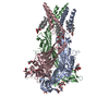 9c2aC  9c2bC M: map data used to model this data C: citing same article ( |
|---|---|
| Similar structure data |
-Links
 PDBj
PDBj- Assembly
| Deposited unit | 
|
|---|---|
| 1 |
|
-Components
| #1: Protein | Mass: 45038.957 Da / Num. of mol.: 3 Source method: isolated from a genetically manipulated source Source: (gene. exp.) Homo sapiens (human) / Gene: P2RX1, P2X1 / Cell (production host): HEK293 GNTI- / Production host: Homo sapiens (human) / References: UniProt: P51575#2: Sugar | ChemComp-NAG /   Type: D-saccharide, beta linking / Mass: 221.208 Da / Num. of mol.: 9 / Source method: obtained synthetically / Formula: C8H15NO6 Type: D-saccharide, beta linking / Mass: 221.208 Da / Num. of mol.: 9 / Source method: obtained synthetically / Formula: C8H15NO6#3: Chemical | ChemComp-A1ALI / Mass: 1329.236 Da / Num. of mol.: 6 / Source method: obtained synthetically / Formula: C41H32N6O29S8 / Feature type: SUBJECT OF INVESTIGATION Has ligand of interest | Y | Has protein modification | Y | |
|---|
-Experimental details
-Experiment
| Experiment | Method: ELECTRON MICROSCOPY |
|---|---|
| EM experiment | Aggregation state: PARTICLE / 3D reconstruction method: single particle reconstruction |
- Sample preparation
| Component | Name: Membrane protein / Type: COMPLEX / Entity ID: #1 / Source: RECOMBINANT |
|---|---|
| Source (natural) | Organism: Homo sapiens (human) |
| Source (recombinant) | Organism: Homo sapiens (human) / Cell: HEK293 GNTI- |
| Buffer solution | pH: 7 |
| Specimen | Embedding applied: NO / Shadowing applied: NO / Staining applied: NO / Vitrification applied: YES |
| Vitrification | Instrument: FEI VITROBOT MARK IV / Cryogen name: ETHANE |
- Electron microscopy imaging
| Experimental equipment |  Model: Titan Krios / Image courtesy: FEI Company |
|---|---|
| Microscopy | Model: FEI TITAN KRIOS |
| Electron gun | Electron source:  FIELD EMISSION GUN / Accelerating voltage: 300 kV / Illumination mode: FLOOD BEAM FIELD EMISSION GUN / Accelerating voltage: 300 kV / Illumination mode: FLOOD BEAM |
| Electron lens | Mode: BRIGHT FIELD / Nominal magnification: 130000 X / Nominal defocus max: 1400 nm / Nominal defocus min: 900 nm / Cs: 2.7 mm / C2 aperture diameter: 70 µm |
| Specimen holder | Specimen holder model: FEI TITAN KRIOS AUTOGRID HOLDER |
| Image recording | Electron dose: 45 e/Å2 / Film or detector model: GATAN K3 (6k x 4k) / Num. of real images: 22866 |
| EM imaging optics | Energyfilter slit width: 20 eV |
- Processing
| EM software | Name: PHENIX / Category: model refinement | ||||||||||||||||||||||||
|---|---|---|---|---|---|---|---|---|---|---|---|---|---|---|---|---|---|---|---|---|---|---|---|---|---|
| CTF correction | Type: PHASE FLIPPING AND AMPLITUDE CORRECTION | ||||||||||||||||||||||||
| Symmetry | Point symmetry: C3 (3 fold cyclic) | ||||||||||||||||||||||||
| 3D reconstruction | Resolution: 2.9 Å / Resolution method: FSC 0.143 CUT-OFF / Num. of particles: 86114 / Symmetry type: POINT | ||||||||||||||||||||||||
| Refine LS restraints |
|
