 Movie
Movie Controller


+ Open data

- Basic information
| Entry | Database: PDB / ID: 9baq | ||||||
|---|---|---|---|---|---|---|---|
| Title | CryoEM structure of DIM2-HP1-H3K9me3-DNA complex | ||||||
 Components Components |
| ||||||
 Keywords Keywords | Transferase/DNA Binding Protein/DNA / DNA methyltransferase / Transferase-DNA Binding Protein-DNA complex | ||||||
| Function / homology |  Function and homology information Function and homology informationDNA (cytosine-5-)-methyltransferase / DNA (cytosine-5-)-methyltransferase activity / negative regulation of gene expression via chromosomal CpG island methylation / heterochromatin / structural constituent of chromatin / nucleosome / methylation / chromatin remodeling / protein heterodimerization activity / chromatin binding ...DNA (cytosine-5-)-methyltransferase / DNA (cytosine-5-)-methyltransferase activity / negative regulation of gene expression via chromosomal CpG island methylation / heterochromatin / structural constituent of chromatin / nucleosome / methylation / chromatin remodeling / protein heterodimerization activity / chromatin binding / DNA binding / nucleus Similarity search - Function | ||||||
| Biological species |  Neurospora crassa (fungus) Neurospora crassa (fungus) | ||||||
| Method | ELECTRON MICROSCOPY / single particle reconstruction / cryo EM / Resolution: 2.79 Å | ||||||
 Authors Authors | Song, J. / Shao, Z. | ||||||
| Funding support |  United States, 1items United States, 1items
| ||||||
 Citation Citation | Journal: Nat Commun / Year: 2024 Title: Multi-layered heterochromatin interaction as a switch for DIM2-mediated DNA methylation. Authors: Zengyu Shao / Jiuwei Lu / Nelli Khudaverdyan / Jikui Song / Abstract: Functional crosstalk between DNA methylation, histone H3 lysine-9 trimethylation (H3K9me3) and heterochromatin protein 1 (HP1) is essential for proper heterochromatin assembly and genome stability. ...Functional crosstalk between DNA methylation, histone H3 lysine-9 trimethylation (H3K9me3) and heterochromatin protein 1 (HP1) is essential for proper heterochromatin assembly and genome stability. However, how repressive chromatin cues guide DNA methyltransferases for region-specific DNA methylation remains largely unknown. Here, we report structure-function characterizations of DNA methyltransferase Defective-In-Methylation-2 (DIM2) in Neurospora. The DNA methylation activity of DIM2 requires the presence of both H3K9me3 and HP1. Our structural study reveals a bipartite DIM2-HP1 interaction, leading to a disorder-to-order transition of the DIM2 target-recognition domain that is essential for substrate binding. Furthermore, the structure of DIM2-HP1-H3K9me3-DNA complex reveals a substrate-binding mechanism distinct from that for its mammalian orthologue DNMT1. In addition, the dual recognition of H3K9me3 peptide by the DIM2 RFTS and BAH1 domains allosterically impacts the DIM2-substrate binding, thereby controlling DIM2-mediated DNA methylation. Together, this study uncovers how multiple heterochromatin factors coordinately orchestrate an activity-switching mechanism for region-specific DNA methylation. | ||||||
| History |
|
- Structure visualization
| Structure viewer | Molecule: MolmilJmol/JSmol |
|---|
- Downloads & links
-Download
| PDBx/mmCIF format | 9baq.cif.gz | 275.1 KB | Display | PDBx/mmCIF format |
|---|---|---|---|---|
| PDB format | pdb9baq.ent.gz | 202.3 KB | Display | PDB format |
| PDBx/mmJSON format | 9baq.json.gz | Tree view | PDBx/mmJSON format | |
| Others |  Other downloads Other downloads |
-Validation report
| Arichive directory | https://data.pdbj.org/pub/pdb/validation_reports/ba/9baqftp://data.pdbj.org/pub/pdb/validation_reports/ba/9baq | HTTPS FTP |
|---|
-Related structure data
| Related structure data | 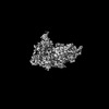 44411MC 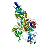 9bapC  9bazC M: map data used to model this data C: citing same article ( |
|---|---|
| Similar structure data |


-Links
 PDBj
PDBj


- Assembly
| Deposited unit | 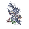
|
|---|---|
| 1 |
|
-Components
-Protein , 2 types, 3 molecules ABC
| #1: Protein | Mass: 139935.891 Da / Num. of mol.: 1 Source method: isolated from a genetically manipulated source Source: (gene. exp.) Neurospora crassa (fungus) / Gene: dim-2, GE21DRAFT_10473 / Production host:  References: UniProt: Q96W73, DNA (cytosine-5-)-methyltransferase |
|---|---|
| #2: Protein | Mass: 30489.086 Da / Num. of mol.: 2 Source method: isolated from a genetically manipulated source Source: (gene. exp.) Neurospora crassa (fungus) / Gene: 49D12.150, hpo, GE21DRAFT_9232 / Production host: |
-Protein/peptide , 1 types, 2 molecules DF
| #3: Protein/peptide | Mass: 2791.302 Da / Num. of mol.: 2 Source method: isolated from a genetically manipulated source Source: (gene. exp.) Neurospora crassa (fungus) / Production host: |
|---|
-DNA chain , 2 types, 2 molecules GH
| #4: DNA chain | Mass: 5709.714 Da / Num. of mol.: 1 Source method: isolated from a genetically manipulated source Source: (gene. exp.) Neurospora crassa (fungus) / Production host: |
|---|---|
| #5: DNA chain | Mass: 5323.441 Da / Num. of mol.: 1 Source method: isolated from a genetically manipulated source Source: (gene. exp.) Neurospora crassa (fungus) / Production host: |
-Non-polymers , 2 types, 2 molecules 

| #6: Chemical | ChemComp-SAH /  Mass: 384.411 Da / Num. of mol.: 1 / Source method: obtained synthetically / Formula: C14H20N6O5S Mass: 384.411 Da / Num. of mol.: 1 / Source method: obtained synthetically / Formula: C14H20N6O5S |
|---|---|
| #7: Chemical | ChemComp-ZN /  Mass: 65.409 Da / Num. of mol.: 1 / Source method: obtained synthetically / Formula: Zn Mass: 65.409 Da / Num. of mol.: 1 / Source method: obtained synthetically / Formula: Zn |
-Details
| Has ligand of interest | N |
|---|---|
| Has protein modification | Y |
-Experimental details
-Experiment
| Experiment | Method: ELECTRON MICROSCOPY |
|---|---|
| EM experiment | Aggregation state: PARTICLE / 3D reconstruction method: single particle reconstruction |
- Sample preparation
| Component | Name: DIM2-HP1-H3K9m3-DNA / Type: COMPLEX / Entity ID: #1-#5 / Source: RECOMBINANT |
|---|---|
| Source (natural) | Organism: Neurospora crassa (fungus) |
| Source (recombinant) | Organism: |
| Buffer solution | pH: 8 |
| Specimen | Embedding applied: NO / Shadowing applied: NO / Staining applied: NO / Vitrification applied: YES |
| Vitrification | Cryogen name: ETHANE |
- Electron microscopy imaging
| Experimental equipment |  Model: Titan Krios / Image courtesy: FEI Company |
|---|---|
| Microscopy | Model: FEI TITAN KRIOS |
| Electron gun | Electron source:  FIELD EMISSION GUN / Accelerating voltage: 300 kV / Illumination mode: FLOOD BEAM FIELD EMISSION GUN / Accelerating voltage: 300 kV / Illumination mode: FLOOD BEAM |
| Electron lens | Mode: BRIGHT FIELD / Nominal defocus max: 2500 nm / Nominal defocus min: 800 nm |
| Image recording | Electron dose: 50 e/Å2 / Film or detector model: GATAN K3 BIOQUANTUM (6k x 4k) |
- Processing
| EM software | Name: PHENIX / Version: 1.21_5207: / Category: model refinement | ||||||||||||||||||||||||
|---|---|---|---|---|---|---|---|---|---|---|---|---|---|---|---|---|---|---|---|---|---|---|---|---|---|
| CTF correction | Type: PHASE FLIPPING AND AMPLITUDE CORRECTION | ||||||||||||||||||||||||
| 3D reconstruction | Resolution: 2.79 Å / Resolution method: FSC 0.143 CUT-OFF / Num. of particles: 128667 / Symmetry type: POINT | ||||||||||||||||||||||||
| Refine LS restraints |
|
