 Movie
Movie Controller

[English] 日本語
 Yorodumi
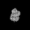
- PDB-9b8t: Human polymerase epsilon bound to PCNA and DNA in the nucleotide ... -
+ Open data

- Basic information
| Entry | Database: PDB / ID: 9b8t | |||||||||
|---|---|---|---|---|---|---|---|---|---|---|
| Title | Human polymerase epsilon bound to PCNA and DNA in the nucleotide bound state | |||||||||
 Components Components |
| |||||||||
 Keywords Keywords | DNA Binding Protein/DNA / DNA polymerase / DNA / DNA Binding Protein-DNA complex | |||||||||
| Function / homology |  Function and homology information Function and homology informationepsilon DNA polymerase complex / dinucleotide insertion or deletion binding / PCNA-p21 complex / mitotic telomere maintenance via semi-conservative replication / purine-specific mismatch base pair DNA N-glycosylase activity / nuclear lamina / Polymerase switching / Processive synthesis on the lagging strand / PCNA complex / MutLalpha complex binding ...epsilon DNA polymerase complex / dinucleotide insertion or deletion binding / PCNA-p21 complex / mitotic telomere maintenance via semi-conservative replication / purine-specific mismatch base pair DNA N-glycosylase activity / nuclear lamina / Polymerase switching / Processive synthesis on the lagging strand / PCNA complex / MutLalpha complex binding / Removal of the Flap Intermediate / Telomere C-strand (Lagging Strand) Synthesis / Mismatch repair (MMR) directed by MSH2:MSH3 (MutSbeta) / Mismatch repair (MMR) directed by MSH2:MSH6 (MutSalpha) / Transcription of E2F targets under negative control by DREAM complex / Polymerase switching on the C-strand of the telomere / replisome / Processive synthesis on the C-strand of the telomere / response to L-glutamate / Removal of the Flap Intermediate from the C-strand / response to dexamethasone / histone acetyltransferase binding / leading strand elongation / DNA polymerase processivity factor activity / G1/S-Specific Transcription / DNA synthesis involved in DNA repair / nuclear replication fork / SUMOylation of DNA replication proteins / replication fork processing / PCNA-Dependent Long Patch Base Excision Repair / response to cadmium ion / estrous cycle / mismatch repair / cyclin-dependent protein kinase holoenzyme complex / base-excision repair, gap-filling / translesion synthesis / epithelial cell differentiation / DNA polymerase binding / TP53 Regulates Transcription of Genes Involved in G2 Cell Cycle Arrest / liver regeneration / positive regulation of DNA replication / nuclear estrogen receptor binding / replication fork / positive regulation of DNA repair / Translesion synthesis by REV1 / Translesion synthesis by POLK / male germ cell nucleus / Translesion synthesis by POLI / Gap-filling DNA repair synthesis and ligation in GG-NER / Termination of translesion DNA synthesis / Translesion Synthesis by POLH / Recognition of DNA damage by PCNA-containing replication complex / receptor tyrosine kinase binding / DNA-templated DNA replication / cellular response to xenobiotic stimulus / HDR through Homologous Recombination (HRR) / Dual Incision in GG-NER / cellular response to hydrogen peroxide / Dual incision in TC-NER / Gap-filling DNA repair synthesis and ligation in TC-NER / cellular response to UV / response to estradiol / E3 ubiquitin ligases ubiquitinate target proteins / heart development / chromatin organization / 4 iron, 4 sulfur cluster binding / DNA-directed DNA polymerase / damaged DNA binding / DNA-directed DNA polymerase activity / chromosome, telomeric region / nuclear body / nucleotide binding / chromatin binding / centrosome / chromatin / protein-containing complex binding / enzyme binding / negative regulation of transcription by RNA polymerase II / DNA binding / extracellular exosome / zinc ion binding / nucleoplasm / identical protein binding / nucleus Similarity search - Function | |||||||||
| Biological species |  Homo sapiens (human) Homo sapiens (human)DNA molecule (others) | |||||||||
| Method | ELECTRON MICROSCOPY / single particle reconstruction / cryo EM / Resolution: 2.95 Å | |||||||||
 Authors Authors | Wang, F. / He, Q. / Li, H. | |||||||||
| Funding support |  United States, 2items United States, 2items
| |||||||||
 Citation Citation | Journal: Nat Commun / Year: 2024 Title: Structures of the human leading strand Polε-PCNA holoenzyme. Authors: Qing He / Feng Wang / Nina Y Yao / Michael E O'Donnell / Huilin Li / Abstract: In eukaryotes, the leading strand DNA is synthesized by Polε and the lagging strand by Polδ. These replicative polymerases have higher processivity when paired with the DNA clamp PCNA. While the ...In eukaryotes, the leading strand DNA is synthesized by Polε and the lagging strand by Polδ. These replicative polymerases have higher processivity when paired with the DNA clamp PCNA. While the structure of the yeast Polε catalytic domain has been determined, how Polε interacts with PCNA is unknown in any eukaryote, human or yeast. Here we report two cryo-EM structures of human Polε-PCNA-DNA complex, one in an incoming nucleotide bound state and the other in a nucleotide exchange state. The structures reveal an unexpected three-point interface between the Polε catalytic domain and PCNA, with the conserved PIP (PCNA interacting peptide)-motif, the unique P-domain, and the thumb domain each interacting with a different protomer of the PCNA trimer. We propose that the multi-point interface prevents other PIP-containing factors from recruiting to PCNA while PCNA functions with Polε. Comparison of the two states reveals that the finger domain pivots around the [4Fe-4S] cluster-containing tip of the P-domain to regulate nucleotide exchange and incoming nucleotide binding. | |||||||||
| History |
|
- Structure visualization
| Structure viewer | Molecule: MolmilJmol/JSmol |
|---|
- Downloads & links
-Download
| PDBx/mmCIF format | 9b8t.cif.gz | 444.4 KB | Display | PDBx/mmCIF format |
|---|---|---|---|---|
| PDB format | pdb9b8t.ent.gz | 338.9 KB | Display | PDB format |
| PDBx/mmJSON format | 9b8t.json.gz | Tree view | PDBx/mmJSON format | |
| Others |  Other downloads Other downloads |
-Validation report
| Arichive directory | https://data.pdbj.org/pub/pdb/validation_reports/b8/9b8tftp://data.pdbj.org/pub/pdb/validation_reports/b8/9b8t | HTTPS FTP |
|---|
-Related structure data
| Related structure data | 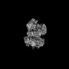 44358MC 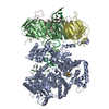 9b8sC M: map data used to model this data C: citing same article ( |
|---|---|
| Similar structure data |
-Links
 PDBj
PDBj


- Assembly
| Deposited unit | 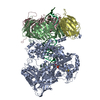
|
|---|---|
| 1 |
|
-Components
-Protein , 2 types, 4 molecules ABCD
| #1: Protein | Mass: 261782.266 Da / Num. of mol.: 1 / Mutation: D275A, E277A Source method: isolated from a genetically manipulated source Source: (gene. exp.) Homo sapiens (human) / Gene: POLE1Production host: Insect cell expression vector pTIE1 (others) References: UniProt: Q9Y5S4, DNA-directed DNA polymerase |
|---|---|
| #2: Protein | Mass: 28795.752 Da / Num. of mol.: 3 Source method: isolated from a genetically manipulated source Source: (gene. exp.) Homo sapiens (human) / Gene: PCNAProduction host: Insect cell expression vector pTIE1 (others) References: UniProt: P12004 |
-DNA chain , 2 types, 2 molecules PT
| #3: DNA chain | Mass: 10823.965 Da / Num. of mol.: 1 / Source method: obtained synthetically / Source: (synth.) DNA molecule (others) |
|---|---|
| #4: DNA chain | Mass: 18136.680 Da / Num. of mol.: 1 / Source method: obtained synthetically / Source: (synth.) DNA molecule (others) |
-Non-polymers , 3 types, 3 molecules 


| #5: Chemical | ChemComp-TTP /  Mass: 482.168 Da / Num. of mol.: 1 / Source method: obtained synthetically / Formula: C10H17N2O14P3 / Feature type: SUBJECT OF INVESTIGATION Mass: 482.168 Da / Num. of mol.: 1 / Source method: obtained synthetically / Formula: C10H17N2O14P3 / Feature type: SUBJECT OF INVESTIGATION |
|---|---|
| #6: Chemical | ChemComp-MG /  Mass: 24.305 Da / Num. of mol.: 1 / Source method: obtained synthetically / Formula: Mg / Feature type: SUBJECT OF INVESTIGATION Mass: 24.305 Da / Num. of mol.: 1 / Source method: obtained synthetically / Formula: Mg / Feature type: SUBJECT OF INVESTIGATION |
| #7: Chemical | ChemComp-SF4 /  Mass: 351.640 Da / Num. of mol.: 1 / Source method: obtained synthetically / Formula: Fe4S4 / Feature type: SUBJECT OF INVESTIGATION Mass: 351.640 Da / Num. of mol.: 1 / Source method: obtained synthetically / Formula: Fe4S4 / Feature type: SUBJECT OF INVESTIGATION |
-Details
| Has ligand of interest | Y |
|---|
-Experimental details
-Experiment
| Experiment | Method: ELECTRON MICROSCOPY |
|---|---|
| EM experiment | Aggregation state: PARTICLE / 3D reconstruction method: single particle reconstruction |
- Sample preparation
| Component | Name: The DNA bound Pol epsilon and PCNA complex / Type: COMPLEX / Entity ID: #1-#4 / Source: RECOMBINANT | ||||||||||||||||||||
|---|---|---|---|---|---|---|---|---|---|---|---|---|---|---|---|---|---|---|---|---|---|
| Molecular weight | Value: 0.45 MDa / Experimental value: YES | ||||||||||||||||||||
| Source (natural) | Organism: Homo sapiens (human) | ||||||||||||||||||||
| Source (recombinant) | Organism: Insect cell expression vector pTIE1 (others) | ||||||||||||||||||||
| Buffer solution | pH: 7.5 | ||||||||||||||||||||
| Buffer component |
| ||||||||||||||||||||
| Specimen | Conc.: 0.75 mg/ml / Embedding applied: NO / Shadowing applied: NO / Staining applied: NO / Vitrification applied: YES | ||||||||||||||||||||
| Specimen support | Grid material: GOLD / Grid mesh size: 400 divisions/in. / Grid type: Quantifoil R1.2/1.3 | ||||||||||||||||||||
| Vitrification | Instrument: FEI VITROBOT MARK IV / Cryogen name: ETHANE / Humidity: 100 % / Chamber temperature: 280 K |
- Electron microscopy imaging
| Experimental equipment |  Model: Titan Krios / Image courtesy: FEI Company |
|---|---|
| Microscopy | Model: FEI TITAN KRIOS |
| Electron gun | Electron source:  FIELD EMISSION GUN / Accelerating voltage: 300 kV / Illumination mode: FLOOD BEAM FIELD EMISSION GUN / Accelerating voltage: 300 kV / Illumination mode: FLOOD BEAM |
| Electron lens | Mode: BRIGHT FIELD / Nominal defocus max: 1800 nm / Nominal defocus min: 1200 nm |
| Image recording | Electron dose: 60 e/Å2 / Film or detector model: GATAN K3 (6k x 4k) |
- Processing
| EM software |
| ||||||||||||||||||||||||
|---|---|---|---|---|---|---|---|---|---|---|---|---|---|---|---|---|---|---|---|---|---|---|---|---|---|
| CTF correction | Type: PHASE FLIPPING AND AMPLITUDE CORRECTION | ||||||||||||||||||||||||
| 3D reconstruction | Resolution: 2.95 Å / Resolution method: FSC 0.143 CUT-OFF / Num. of particles: 320111 / Symmetry type: POINT | ||||||||||||||||||||||||
| Atomic model building | Protocol: RIGID BODY FIT / Space: REAL | ||||||||||||||||||||||||
| Refine LS restraints |
|
