 Movie
Movie Controller


+ Open data

- Basic information
| Entry | Database: PDB / ID: 9b1m | ||||||
|---|---|---|---|---|---|---|---|
| Title | Pyrazinoate bound human URAT1 in the inward-facing state (site1) | ||||||
 Components Components | Solute carrier family 22 member 12 | ||||||
 Keywords Keywords | MEMBRANE PROTEIN / SLC transporter / SLC22A family / uric acid / inward facing | ||||||
| Function / homology |  Function and homology information Function and homology informationDefective SLC22A12 causes renal hypouricemia 1 (RHUC1) / Organic anion transport by SLC22 transporters / renal urate salt excretion / urate transport / urate metabolic process / urate transmembrane transporter activity / : / cellular homeostasis / monoatomic ion transport / PDZ domain binding ...Defective SLC22A12 causes renal hypouricemia 1 (RHUC1) / Organic anion transport by SLC22 transporters / renal urate salt excretion / urate transport / urate metabolic process / urate transmembrane transporter activity / : / cellular homeostasis / monoatomic ion transport / PDZ domain binding / brush border membrane / cellular response to insulin stimulus / apical plasma membrane / response to xenobiotic stimulus / extracellular exosome / membrane / plasma membrane Similarity search - Function | ||||||
| Biological species |  Homo sapiens (human) Homo sapiens (human) | ||||||
| Method | ELECTRON MICROSCOPY / single particle reconstruction / cryo EM / Resolution: 3 Å | ||||||
 Authors Authors | Dai, Y. / Lee, C.H. | ||||||
| Funding support |  United States, 1items United States, 1items
| ||||||
 Citation Citation | Journal: Cell Res / Year: 2024 Title: Transport mechanism and structural pharmacology of human urate transporter URAT1. Authors: Yaxin Dai / Chia-Hsueh Lee / Abstract: Urate is an endogenous product of purine metabolism in the liver. High urate levels in the blood lead to gout, a very common and painful inflammatory arthritis. Excreted urate is reabsorbed in the ...Urate is an endogenous product of purine metabolism in the liver. High urate levels in the blood lead to gout, a very common and painful inflammatory arthritis. Excreted urate is reabsorbed in the kidney mainly by URAT1 antiporter, a key target for anti-gout drugs. To uncover the mechanisms of urate transport and drug inhibition, we determined cryo-EM structures of human URAT1 with urate, counter anion pyrazinoate, or anti-gout drugs of different chemotypes - lesinurad, verinurad, and dotinurad. We captured the outward-to-inward transition of URAT1 during urate uptake, revealing that urate binds in a phenylalanine-rich pocket and engages with key gating residues to drive the transport cycle. In contrast to the single binding site for urate, pyrazinoate interacts with three distinct, functionally relevant sites within URAT1, a mechanism that has not yet been observed in other anion antiporters. In addition, we found that while all three drugs compete with substrates and halt the transport cycle, verinurad and dotinurad further hijack gating residues to achieve high potency. These insights advance our understanding of organic anion transport and provide a foundation for designing improved gout therapeutics. | ||||||
| History |
|
- Structure visualization
| Structure viewer | Molecule: MolmilJmol/JSmol |
|---|
- Downloads & links
-Download
| PDBx/mmCIF format | 9b1m.cif.gz | 111.2 KB | Display | PDBx/mmCIF format |
|---|---|---|---|---|
| PDB format | pdb9b1m.ent.gz | 83.3 KB | Display | PDB format |
| PDBx/mmJSON format | 9b1m.json.gz | Tree view | PDBx/mmJSON format | |
| Others |  Other downloads Other downloads |
-Validation report
| Arichive directory | https://data.pdbj.org/pub/pdb/validation_reports/b1/9b1mftp://data.pdbj.org/pub/pdb/validation_reports/b1/9b1m | HTTPS FTP |
|---|
-Related structure data
| Related structure data | 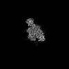 44084MC 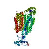 9b1fC 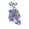 9b1gC 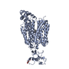 9b1hC 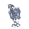 9b1iC 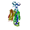 9b1jC 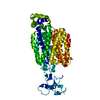 9b1kC 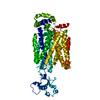 9b1lC 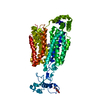 9b1nC 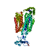 9b1oC M: map data used to model this data C: citing same article ( |
|---|---|
| Similar structure data |
-Links
 PDBj
PDBj


- Assembly
| Deposited unit | 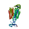
|
|---|---|
| 1 |
|
-Components
| #1: Protein | Mass: 59257.402 Da / Num. of mol.: 1 Source method: isolated from a genetically manipulated source Source: (gene. exp.) Homo sapiens (human) / Gene: SLC22A12, OATL4, URAT1, UNQ6453/PRO34004 / Production host: Homo sapiens (human) / References: UniProt: Q96S37 |
|---|---|
| #2: Sugar | ChemComp-NAG /   Type: D-saccharide, beta linking / Mass: 221.208 Da / Num. of mol.: 1 Type: D-saccharide, beta linking / Mass: 221.208 Da / Num. of mol.: 1Source method: isolated from a genetically manipulated source Formula: C8H15NO6 |
| #3: Chemical | ChemComp-VGL / 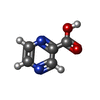  Mass: 124.097 Da / Num. of mol.: 1 / Source method: obtained synthetically / Formula: C5H4N2O2 / Feature type: SUBJECT OF INVESTIGATION Mass: 124.097 Da / Num. of mol.: 1 / Source method: obtained synthetically / Formula: C5H4N2O2 / Feature type: SUBJECT OF INVESTIGATION |
| Has ligand of interest | Y |
| Has protein modification | Y |
-Experimental details
-Experiment
| Experiment | Method: ELECTRON MICROSCOPY |
|---|---|
| EM experiment | Aggregation state: PARTICLE / 3D reconstruction method: single particle reconstruction |
- Sample preparation
| Component | Name: Pyrazinoate bound human URAT1 in the inward-facing state (site1). Type: COMPLEX / Entity ID: #1 / Source: RECOMBINANT |
|---|---|
| Source (natural) | Organism: Homo sapiens (human) |
| Source (recombinant) | Organism: Homo sapiens (human) |
| Buffer solution | pH: 6.5 |
| Specimen | Embedding applied: NO / Shadowing applied: NO / Staining applied: NO / Vitrification applied: YES |
| Vitrification | Cryogen name: ETHANE |
- Electron microscopy imaging
| Experimental equipment |  Model: Titan Krios / Image courtesy: FEI Company |
|---|---|
| Microscopy | Model: FEI TITAN KRIOS |
| Electron gun | Electron source:  FIELD EMISSION GUN / Accelerating voltage: 300 kV / Illumination mode: OTHER FIELD EMISSION GUN / Accelerating voltage: 300 kV / Illumination mode: OTHER |
| Electron lens | Mode: BRIGHT FIELD / Nominal defocus max: 2000 nm / Nominal defocus min: 1000 nm / Cs: 2.7 mm |
| Image recording | Electron dose: 68.9 e/Å2 / Film or detector model: GATAN K3 (6k x 4k) |
- Processing
| EM software | Name: PHENIX / Version: 1.21_5207: / Category: model refinement |
|---|---|
| CTF correction | Type: PHASE FLIPPING AND AMPLITUDE CORRECTION |
| 3D reconstruction | Resolution: 3 Å / Resolution method: FSC 0.143 CUT-OFF / Num. of particles: 55197 / Symmetry type: POINT |
