 Movie
Movie Controller

[English] 日本語
 Yorodumi
- PDB-8ys2: Overall structure of Eastern Equine Encephalitis virus VLP in com... -
+ Open data

- Basic information
| Entry | Database: PDB / ID: 8ys2 | ||||||||||||
|---|---|---|---|---|---|---|---|---|---|---|---|---|---|
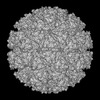
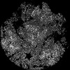
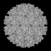
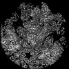
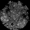
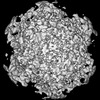
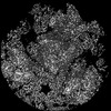
| Title | Overall structure of Eastern Equine Encephalitis virus VLP in complex with the receptor VLDLR LA1-2 | ||||||||||||
 Components Components |
| ||||||||||||
 Keywords Keywords | VIRUS LIKE PARTICLE / Eastern Equine Encephalitis virus / EEEV / receptor / complex / VLDLR / glycoprotein / VIRAL PROTEIN | ||||||||||||
| Function / homology |  Function and homology information Function and homology informationreelin receptor activity / VLDL clearance / glycoprotein transport / ventral spinal cord development / very-low-density lipoprotein particle receptor activity / Reelin signalling pathway / very-low-density lipoprotein particle binding / reelin-mediated signaling pathway / low-density lipoprotein particle receptor activity / togavirin ...reelin receptor activity / VLDL clearance / glycoprotein transport / ventral spinal cord development / very-low-density lipoprotein particle receptor activity / Reelin signalling pathway / very-low-density lipoprotein particle binding / reelin-mediated signaling pathway / low-density lipoprotein particle receptor activity / togavirin / very-low-density lipoprotein particle clearance / very-low-density lipoprotein particle / T=4 icosahedral viral capsid / positive regulation of dendrite development / dendrite morphogenesis / cargo receptor activity / lipid transport / apolipoprotein binding / clathrin-coated pit / cholesterol metabolic process / VLDLR internalisation and degradation / receptor-mediated endocytosis / memory / symbiont-mediated suppression of host gene expression / calcium-dependent protein binding / nervous system development / symbiont-mediated suppression of host toll-like receptor signaling pathway / host cell cytoplasm / receptor complex / symbiont entry into host cell / lysosomal membrane / serine-type endopeptidase activity / fusion of virus membrane with host endosome membrane / calcium ion binding / host cell nucleus / virion attachment to host cell / host cell plasma membrane / virion membrane / structural molecule activity / signal transduction / proteolysis / RNA binding / membrane / plasma membrane Similarity search - Function | ||||||||||||
| Biological species |  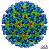 Eastern equine encephalitis virus Eastern equine encephalitis virus Homo sapiens (human) Homo sapiens (human) | ||||||||||||
| Method | ELECTRON MICROSCOPY / single particle reconstruction / cryo EM / Resolution: 5.2 Å | ||||||||||||
 Authors Authors | Cao, D. / Ma, B. / Cao, Z. / Xu, X. / Zhang, X. / Xiang, Y. | ||||||||||||
| Funding support |  China, 3items China, 3items
| ||||||||||||
 Citation Citation | Journal: Nat Commun / Year: 2024 Title: The receptor VLDLR binds Eastern Equine Encephalitis virus through multiple distinct modes. Authors: Duanfang Cao / Bingting Ma / Ziyi Cao / Xiaoyu Xu / Xinzheng Zhang / Ye Xiang / Abstract: Eastern Equine Encephalitis virus (EEEV) is an alphavirus that can cause severe diseases in infected humans. The very low-density lipoprotein receptor (VLDLR) was recently identified as a receptor of ...Eastern Equine Encephalitis virus (EEEV) is an alphavirus that can cause severe diseases in infected humans. The very low-density lipoprotein receptor (VLDLR) was recently identified as a receptor of EEEV. Herein, we performed cryo-electron microscopy structural and biochemistry studies on the specific interactions between EEEV and VLDLR. Our results show that VLDLR binds EEEV at three different sites A, B and C through its membrane-distal LDLR class A (LA) repeats. Site A is located in the cleft in between the E1-E2 heterodimers. Site B is located near the connecting β ribbon of E2 and is in proximity to site A, while site C is on the domain B of E2. The binding of VLDLR LAs to EEEV is in complex modes, including the LA1-2 and LA3-5 mediated two major modes. Disruption of the LA1-2 mediated binding significantly affect the cell attachment of EEEV. However, the mutation W132G of VLDLR impairs the binding of LA3, drives the switch of the binding modes, and significantly enhances the attachment of EEEV to the cell. The W132G variant of VLDLR could be identified in human genome and SNP sequences, implying that people with similar mutations in VLDLR may be highly susceptible to EEEV infection. | ||||||||||||
| History |
|
- Structure visualization
| Structure viewer | Molecule: MolmilJmol/JSmol |
|---|
- Downloads & links
-Download
| PDBx/mmCIF format | 8ys2.cif.gz | 835.6 KB | Display | PDBx/mmCIF format |
|---|---|---|---|---|
| PDB format | pdb8ys2.ent.gz | Display | PDB format | |
| PDBx/mmJSON format | 8ys2.json.gz | Tree view | PDBx/mmJSON format | |
| Others |  Other downloads Other downloads |
-Validation report
| Summary document | 8ys2_validation.pdf.gz | 1.7 MB | Display | wwPDB validaton report |
|---|---|---|---|---|
| Full document | 8ys2_full_validation.pdf.gz | 1.7 MB | Display | |
| Data in XML | 8ys2_validation.xml.gz | 121.9 KB | Display | |
| Data in CIF | 8ys2_validation.cif.gz | 182.7 KB | Display | |
| Arichive directory | https://data.pdbj.org/pub/pdb/validation_reports/ys/8ys2ftp://data.pdbj.org/pub/pdb/validation_reports/ys/8ys2 | HTTPS FTP |
-Related structure data
| Related structure data | 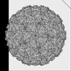 38376MC 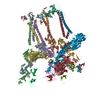 8xi4C  8xi5C 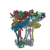 8ys4C M: map data used to model this data C: citing same article ( |
|---|---|
| Similar structure data |

-Links
 PDBj
PDBj


- Assembly
| Deposited unit | 
|
|---|---|
| 1 | x 60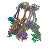
|
-Components
| #1: Protein | Mass: 47984.246 Da / Num. of mol.: 4 Source method: isolated from a genetically manipulated source Source: (gene. exp.) Eastern equine encephalitis virus / Production host: Homo sapiens (human) / References: UniProt: Q4QXJ7#2: Protein | Mass: 47047.020 Da / Num. of mol.: 4 Source method: isolated from a genetically manipulated source Source: (gene. exp.) Eastern equine encephalitis virus / Production host: Homo sapiens (human) / References: UniProt: Q4QXJ7#3: Protein | Mass: 29178.846 Da / Num. of mol.: 4 / Mutation: K67N Source method: isolated from a genetically manipulated source Details: AAU95735.1 / Source: (gene. exp.) Eastern equine encephalitis virus / Production host: Homo sapiens (human) / References: UniProt: Q4QXJ7, togavirin#4: Protein | Mass: 8854.691 Da / Num. of mol.: 4 Source method: isolated from a genetically manipulated source Source: (gene. exp.) Homo sapiens (human) / Gene: VLDLR / Production host: Homo sapiens (human) / References: UniProt: P98155#5: Chemical | ChemComp-CA /   Mass: 40.078 Da / Num. of mol.: 8 / Source method: obtained synthetically / Formula: Ca Mass: 40.078 Da / Num. of mol.: 8 / Source method: obtained synthetically / Formula: CaHas ligand of interest | N | |
|---|
-Experimental details
-Experiment
| Experiment | Method: ELECTRON MICROSCOPY |
|---|---|
| EM experiment | Aggregation state: PARTICLE / 3D reconstruction method: single particle reconstruction |
- Sample preparation
| Component | Name: Eastern equine encephalitis virus / Type: VIRUS Details: EEEV Virus like particle in complexed with its receptor VLDLR LA1-2 Entity ID: #1-#4 / Source: RECOMBINANT |
|---|---|
| Source (natural) | Organism: Eastern equine encephalitis virus |
| Source (recombinant) | Organism: Homo sapiens (human) |
| Details of virus | Empty: YES / Enveloped: YES / Isolate: OTHER / Type: VIRUS-LIKE PARTICLE |
| Natural host | Organism: Eastern equine encephalitis virus |
| Buffer solution | pH: 8 |
| Specimen | Embedding applied: NO / Shadowing applied: NO / Staining applied: NO / Vitrification applied: YES |
| Vitrification | Cryogen name: ETHANE |
- Electron microscopy imaging
| Experimental equipment |  Model: Titan Krios / Image courtesy: FEI Company |
|---|---|
| Microscopy | Model: FEI TITAN KRIOS |
| Electron gun | Electron source:  FIELD EMISSION GUN / Accelerating voltage: 300 kV / Illumination mode: FLOOD BEAM FIELD EMISSION GUN / Accelerating voltage: 300 kV / Illumination mode: FLOOD BEAM |
| Electron lens | Mode: BRIGHT FIELD / Nominal defocus max: 1700 nm / Nominal defocus min: 1200 nm |
| Image recording | Electron dose: 50 e/Å2 / Film or detector model: GATAN K2 SUMMIT (4k x 4k) |
- Processing
| CTF correction | Type: PHASE FLIPPING AND AMPLITUDE CORRECTION | ||||||||||||||||||||||||
|---|---|---|---|---|---|---|---|---|---|---|---|---|---|---|---|---|---|---|---|---|---|---|---|---|---|
| 3D reconstruction | Resolution: 5.2 Å / Resolution method: FSC 0.143 CUT-OFF / Num. of particles: 15654 / Symmetry type: POINT | ||||||||||||||||||||||||
| Refine LS restraints |
|
