 Movie
Movie Controller

[English] 日本語
 Yorodumi
- PDB-8wxb: Cryo-EM structure of the alpha-carboxysome shell vertex from Proc... -
+ Open data

- Basic information
| Entry | Database: PDB / ID: 8wxb | ||||||||||||||||||
|---|---|---|---|---|---|---|---|---|---|---|---|---|---|---|---|---|---|---|---|
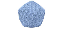
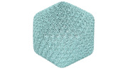
| Title | Cryo-EM structure of the alpha-carboxysome shell vertex from Prochlorococcus MED4 | ||||||||||||||||||
 Components Components |
| ||||||||||||||||||
 Keywords Keywords | PHOTOSYNTHESIS / alpha-carboxysome / carbon fixation | ||||||||||||||||||
| Function / homology |  Function and homology information Function and homology informationstructural constituent of carboxysome shell / carboxysome / carbon fixation / photosynthesis Similarity search - Function | ||||||||||||||||||
| Biological species |  Prochlorococcus sp. MED4 (bacteria) Prochlorococcus sp. MED4 (bacteria) | ||||||||||||||||||
| Method | ELECTRON MICROSCOPY / single particle reconstruction / cryo EM / Resolution: 4.2 Å | ||||||||||||||||||
 Authors Authors | Jiang, Y.L. / Zhou, R.Q. / Zhou, C.Z. / Zeng, Q.L. | ||||||||||||||||||
| Funding support |  China, Hong Kong, 5items China, Hong Kong, 5items
| ||||||||||||||||||
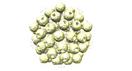
 Citation Citation | Journal: Nat Plants / Year: 2024 Title: Structure and assembly of the α-carboxysome in the marine cyanobacterium Prochlorococcus. Authors: Rui-Qian Zhou / Yong-Liang Jiang / Haofu Li / Pu Hou / Wen-Wen Kong / Jia-Xin Deng / Yuxing Chen / Cong-Zhao Zhou / Qinglu Zeng / Abstract: Carboxysomes are bacterial microcompartments that encapsulate the enzymes RuBisCO and carbonic anhydrase in a proteinaceous shell to enhance the efficiency of photosynthetic carbon fixation. The self- ...Carboxysomes are bacterial microcompartments that encapsulate the enzymes RuBisCO and carbonic anhydrase in a proteinaceous shell to enhance the efficiency of photosynthetic carbon fixation. The self-assembly principles of the intact carboxysome remain elusive. Here we purified α-carboxysomes from Prochlorococcus and examined their intact structures using single-particle cryo-electron microscopy to solve the basic principles of their shell construction and internal RuBisCO organization. The 4.2 Å icosahedral-like shell structure reveals 24 CsoS1 hexamers on each facet and one CsoS4A pentamer at each vertex. RuBisCOs are organized into three concentric layers within the shell, consisting of 72, 32 and up to 4 RuBisCOs at the outer, middle and inner layers, respectively. We uniquely show how full-length and shorter forms of the scaffolding protein CsoS2 bind to the inner surface of the shell via repetitive motifs in the middle and C-terminal regions. Combined with previous reports, we propose a concomitant 'outside-in' assembly principle of α-carboxysomes: the inner surface of the self-assembled shell is reinforced by the middle and C-terminal motifs of the scaffolding protein, while the free N-terminal motifs cluster to recruit RuBisCO in concentric, three-layered spherical arrangements. These new insights into the coordinated assembly of α-carboxysomes may guide the rational design and repurposing of carboxysome structures for improving plant photosynthetic efficiency. | ||||||||||||||||||
| History |
|
- Structure visualization
| Structure viewer | Molecule: MolmilJmol/JSmol |
|---|
- Downloads & links
-Download
| PDBx/mmCIF format | 8wxb.cif.gz | 802.3 KB | Display | PDBx/mmCIF format |
|---|---|---|---|---|
| PDB format | pdb8wxb.ent.gz | 672.1 KB | Display | PDB format |
| PDBx/mmJSON format | 8wxb.json.gz | Tree view | PDBx/mmJSON format | |
| Others |  Other downloads Other downloads |
-Validation report
| Summary document | 8wxb_validation.pdf.gz | 1.6 MB | Display | wwPDB validaton report |
|---|---|---|---|---|
| Full document | 8wxb_full_validation.pdf.gz | 1.6 MB | Display | |
| Data in XML | 8wxb_validation.xml.gz | 135.2 KB | Display | |
| Data in CIF | 8wxb_validation.cif.gz | 213.8 KB | Display | |
| Arichive directory | https://data.pdbj.org/pub/pdb/validation_reports/wx/8wxbftp://data.pdbj.org/pub/pdb/validation_reports/wx/8wxb | HTTPS FTP |
-Related structure data
| Related structure data |  37902MC M: map data used to model this data C: citing same article ( |
|---|---|
| Similar structure data |
-Links
 PDBj
PDBj

- Assembly
| Deposited unit | 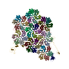
|
|---|---|
| 1 | x 5
|
| 2 |
|
| 3 |
|
| Symmetry | Point symmetry: (Schoenflies symbol: C5 (5 fold cyclic)) |
-Components
| #1: Protein | Mass: 9407.823 Da / Num. of mol.: 1 / Source method: isolated from a natural source / Source: (natural) Prochlorococcus sp. MED4 (bacteria) / References: UniProt: Q7V2C6 | ||
|---|---|---|---|
| #2: Protein | Mass: 10050.501 Da / Num. of mol.: 48 / Source method: isolated from a natural source / Source: (natural) Prochlorococcus sp. MED4 (bacteria) / References: UniProt: Q7V2D1#3: Protein | Mass: 82006.797 Da / Num. of mol.: 2 / Source method: isolated from a natural source / Source: (natural) Prochlorococcus sp. MED4 (bacteria) / References: UniProt: Q7V2C8 |
-Experimental details
-Experiment
| Experiment | Method: ELECTRON MICROSCOPY |
|---|---|
| EM experiment | Aggregation state: PARTICLE / 3D reconstruction method: single particle reconstruction |
- Sample preparation
| Component | Name: The shell vertex of alpha-carboxysome / Type: COMPLEX / Entity ID: all / Source: NATURAL |
|---|---|
| Molecular weight | Value: 3.4 MDa / Experimental value: NO |
| Source (natural) | Organism: Prochlorococcus sp. MED4 (bacteria) |
| Buffer solution | pH: 8.5 |
| Specimen | Embedding applied: NO / Shadowing applied: NO / Staining applied: NO / Vitrification applied: YES |
| Specimen support | Grid type: Quantifoil R2/1 |
| Vitrification | Cryogen name: ETHANE |
- Electron microscopy imaging
| Experimental equipment |  Model: Titan Krios / Image courtesy: FEI Company |
|---|---|
| Microscopy | Model: FEI TITAN KRIOS |
| Electron gun | Electron source:  FIELD EMISSION GUN / Accelerating voltage: 300 kV / Illumination mode: FLOOD BEAM FIELD EMISSION GUN / Accelerating voltage: 300 kV / Illumination mode: FLOOD BEAM |
| Electron lens | Mode: BRIGHT FIELD / Nominal defocus max: 2000 nm / Nominal defocus min: 1000 nm |
| Image recording | Electron dose: 50 e/Å2 / Film or detector model: GATAN K3 (6k x 4k) |
- Processing
| CTF correction | Type: NONE |
|---|---|
| 3D reconstruction | Resolution: 4.2 Å / Resolution method: FSC 0.143 CUT-OFF / Num. of particles: 28093 / Symmetry type: POINT |
