 Movie
Movie Controller


+ Open data

- Basic information
| Entry | Database: PDB / ID: 8wjq | ||||||
|---|---|---|---|---|---|---|---|
| Title | Cryo-EM structure of URAT1(R477S)-Urate complex | ||||||
 Components Components | Solute carrier family 22 member 12 | ||||||
 Keywords Keywords | TRANSPORT PROTEIN / SLC | ||||||
| Function / homology |  Function and homology information Function and homology informationDefective SLC22A12 causes renal hypouricemia 1 (RHUC1) / Organic anion transport / urate transport / renal urate salt excretion / urate transmembrane transporter activity / urate metabolic process / organic anion transport / cellular homeostasis / monoatomic ion transport / PDZ domain binding ...Defective SLC22A12 causes renal hypouricemia 1 (RHUC1) / Organic anion transport / urate transport / renal urate salt excretion / urate transmembrane transporter activity / urate metabolic process / organic anion transport / cellular homeostasis / monoatomic ion transport / PDZ domain binding / brush border membrane / cellular response to insulin stimulus / response to xenobiotic stimulus / apical plasma membrane / extracellular exosome / membrane / plasma membrane Similarity search - Function | ||||||
| Biological species |  Homo sapiens (human) Homo sapiens (human) | ||||||
| Method | ELECTRON MICROSCOPY / single particle reconstruction / cryo EM / Resolution: 3.8 Å | ||||||
 Authors Authors | Qian, H.W. / He, J.J. | ||||||
| Funding support | 1items
| ||||||
 Citation Citation | Journal: Cell Rep / Year: 2024 Title: Structural basis for the transport and substrate selection of human urate transporter 1. Authors: Jingjing He / Guoyun Liu / Fang Kong / Qiulong Tan / Zhenzhou Wang / Meng Yang / Yonglin He / Xiaoxiao Jia / Chuangye Yan / Chao Wang / Hongwu Qian /  Abstract: High serum urate levels are the major risk factor for gout. URAT1, the primary transporter for urate absorption in the kidneys, is well known as an anti-hyperuricemia drug target. However, the ...High serum urate levels are the major risk factor for gout. URAT1, the primary transporter for urate absorption in the kidneys, is well known as an anti-hyperuricemia drug target. However, the clinical application of URAT1-targeted drugs is limited because of their low specificity and severe side effects. The lack of structural information impedes elucidation of the transport mechanism and the development of new drugs. Here, we present the cryoelectron microscopy (cryo-EM) structures of human URAT1(R477S), its complex with urate, and its closely related homolog OAT4. URAT1(R477S) and OAT4 exhibit major facilitator superfamily (MFS) folds with outward- and inward-open conformations, respectively. Structural comparison reveals a 30° rotation between the N-terminal and C-terminal domains, supporting an alternating access mechanism. A conserved arginine (OAT4-Arg473/URAT1-Arg477) is found to be essential for chloride-mediated inhibition. The URAT1(R477S)-urate complex reveals the specificity of urate recognition. Taken together, our study promotes our understanding of the transport mechanism and substrate selection of URAT1. | ||||||
| History |
|
- Structure visualization
| Structure viewer | Molecule: MolmilJmol/JSmol |
|---|
- Downloads & links
-Download
| PDBx/mmCIF format | 8wjq.cif.gz | 100.9 KB | Display | PDBx/mmCIF format |
|---|---|---|---|---|
| PDB format | pdb8wjq.ent.gz | 75.3 KB | Display | PDB format |
| PDBx/mmJSON format | 8wjq.json.gz | Tree view | PDBx/mmJSON format | |
| Others |  Other downloads Other downloads |
-Validation report
| Summary document | 8wjq_validation.pdf.gz | 1.2 MB | Display | wwPDB validaton report |
|---|---|---|---|---|
| Full document | 8wjq_full_validation.pdf.gz | 1.2 MB | Display | |
| Data in XML | 8wjq_validation.xml.gz | 26.1 KB | Display | |
| Data in CIF | 8wjq_validation.cif.gz | 36.3 KB | Display | |
| Arichive directory | https://data.pdbj.org/pub/pdb/validation_reports/wj/8wjqftp://data.pdbj.org/pub/pdb/validation_reports/wj/8wjq | HTTPS FTP |
-Related structure data
| Related structure data |  37589MC 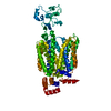 8wjgC 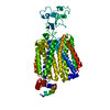 8wjhC M: map data used to model this data C: citing same article ( |
|---|---|
| Similar structure data |

-Links
 PDBj
PDBj


- Assembly
| Deposited unit | 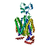
|
|---|---|
| 1 |
|
-Components
| #1: Protein | Mass: 58027.961 Da / Num. of mol.: 1 Source method: isolated from a genetically manipulated source Source: (gene. exp.) Homo sapiens (human) / Gene: SLC22A12, OATL4, URAT1, UNQ6453/PRO34004 / Production host: Homo sapiens (human) / References: UniProt: Q96S37 |
|---|---|
| #2: Chemical | ChemComp-URC /   Mass: 168.110 Da / Num. of mol.: 1 / Source method: obtained synthetically / Formula: C5H4N4O3 / Feature type: SUBJECT OF INVESTIGATION Mass: 168.110 Da / Num. of mol.: 1 / Source method: obtained synthetically / Formula: C5H4N4O3 / Feature type: SUBJECT OF INVESTIGATION |
| Has ligand of interest | Y |
-Experimental details
-Experiment
| Experiment | Method: ELECTRON MICROSCOPY |
|---|---|
| EM experiment | Aggregation state: PARTICLE / 3D reconstruction method: single particle reconstruction |
- Sample preparation
| Component | Name: Cryo-EM structure of URAT1(R477S)-Urate complex / Type: COMPLEX / Entity ID: #1 / Source: RECOMBINANT |
|---|---|
| Source (natural) | Organism: Homo sapiens (human) |
| Buffer solution | pH: 8 |
| Specimen | Embedding applied: NO / Shadowing applied: NO / Staining applied: NO / Vitrification applied: YES |
| Vitrification | Cryogen name: ETHANE |
- Electron microscopy imaging
| Experimental equipment |  Model: Titan Krios / Image courtesy: FEI Company |
|---|---|
| Microscopy | Model: FEI TITAN KRIOS |
| Electron gun | Electron source:  FIELD EMISSION GUN / Accelerating voltage: 300 kV / Illumination mode: FLOOD BEAM FIELD EMISSION GUN / Accelerating voltage: 300 kV / Illumination mode: FLOOD BEAM |
| Electron lens | Mode: BRIGHT FIELD / Nominal defocus max: 1700 nm / Nominal defocus min: 1500 nm |
| Image recording | Electron dose: 50 e/Å2 / Film or detector model: GATAN K3 BIOQUANTUM (6k x 4k) |
- Processing
| CTF correction | Type: NONE | ||||||||||||||||||||||||
|---|---|---|---|---|---|---|---|---|---|---|---|---|---|---|---|---|---|---|---|---|---|---|---|---|---|
| 3D reconstruction | Resolution: 3.8 Å / Resolution method: FSC 0.143 CUT-OFF / Num. of particles: 392817 / Symmetry type: POINT | ||||||||||||||||||||||||
| Refine LS restraints |
|
