 Movie
Movie Controller

[English] 日本語
 Yorodumi
- PDB-8vk4: Structure of mouse RyR1 in complex with S100A1 (high-Ca2+/CFF/ATP... -
+ Open data

- Basic information
| Entry | Database: PDB / ID: 8vk4 | ||||||
|---|---|---|---|---|---|---|---|
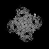
| Title | Structure of mouse RyR1 in complex with S100A1 (high-Ca2+/CFF/ATP dataset) | ||||||
 Components Components |
| ||||||
 Keywords Keywords | MEMBRANE PROTEIN / Calcium / Ion Channel | ||||||
| Function / homology |  Function and homology information Function and homology informationjunctional membrane complex / TGF-beta receptor signaling activates SMADs / Calcineurin activates NFAT / mTORC1-mediated signalling / regulation of response to osmotic stress / sarcoplasmic reticulum calcium ion transport / cytoplasmic side of membrane / regulation of muscle contraction / transforming growth factor beta receptor binding / Stimuli-sensing channels ...junctional membrane complex / TGF-beta receptor signaling activates SMADs / Calcineurin activates NFAT / mTORC1-mediated signalling / regulation of response to osmotic stress / sarcoplasmic reticulum calcium ion transport / cytoplasmic side of membrane / regulation of muscle contraction / transforming growth factor beta receptor binding / Stimuli-sensing channels / type I transforming growth factor beta receptor binding / Ion homeostasis / heart trabecula formation / terminal cisterna / ryanodine receptor complex / S100 protein binding / ryanodine-sensitive calcium-release channel activity / release of sequestered calcium ion into cytosol by sarcoplasmic reticulum / response to caffeine / ossification involved in bone maturation / ventricular cardiac muscle tissue morphogenesis / cellular response to ATP / skin development / regulation of heart contraction / FK506 binding / organelle membrane / positive regulation of sprouting angiogenesis / cellular response to caffeine / outflow tract morphogenesis / extrinsic component of cytoplasmic side of plasma membrane / regulation of ryanodine-sensitive calcium-release channel activity / voltage-gated calcium channel activity / smooth endoplasmic reticulum / skeletal muscle fiber development / heart morphogenesis / T cell proliferation / striated muscle contraction / muscle contraction / release of sequestered calcium ion into cytosol / axon terminus / regulation of cytosolic calcium ion concentration / sarcoplasmic reticulum membrane / T-tubule / calcium channel complex / Hsp70 protein binding / cellular response to calcium ion / sarcomere / sarcoplasmic reticulum / peptidylprolyl isomerase / peptidyl-prolyl cis-trans isomerase activity / negative regulation of transforming growth factor beta receptor signaling pathway / calcium channel activity / sarcolemma / cytokine-mediated signaling pathway / cytoplasmic side of plasma membrane / Z disc / calcium ion transport / ATPase binding / protease binding / protein homotetramerization / vesicle / transmembrane transporter binding / calmodulin binding / calcium ion binding / synapse / perinuclear region of cytoplasm / Golgi apparatus / enzyme binding / protein homodimerization activity / protein-containing complex / mitochondrion / nucleoplasm / ATP binding / identical protein binding / membrane / cytosol / cytoplasm Similarity search - Function | ||||||
| Biological species |  | ||||||
| Method | ELECTRON MICROSCOPY / single particle reconstruction / cryo EM / Resolution: 3.56 Å | ||||||
 Authors Authors | Weninger, G. / Marks, A.R. | ||||||
| Funding support |  United States, 1items United States, 1items
| ||||||
 Citation Citation | Journal: Proc Natl Acad Sci U S A / Year: 2024 Title: Structural insights into the regulation of RyR1 by S100A1. Authors: Gunnar Weninger / Marco C Miotto / Carl Tchagou / Steven Reiken / Haikel Dridi / Sören Brandenburg / Gabriel C Riedemann / Qi Yuan / Yang Liu / Alexander Chang / Anetta Wronska / Stephan E ...Authors: Gunnar Weninger / Marco C Miotto / Carl Tchagou / Steven Reiken / Haikel Dridi / Sören Brandenburg / Gabriel C Riedemann / Qi Yuan / Yang Liu / Alexander Chang / Anetta Wronska / Stephan E Lehnart / Andrew R Marks /  Abstract: S100A1, a small homodimeric EF-hand Ca-binding protein (~21 kDa), plays an important regulatory role in Ca signaling pathways involved in various biological functions including Ca cycling and ...S100A1, a small homodimeric EF-hand Ca-binding protein (~21 kDa), plays an important regulatory role in Ca signaling pathways involved in various biological functions including Ca cycling and contractile performance in skeletal and cardiac myocytes. One key target of the S100A1 interactome is the ryanodine receptor (RyR), a huge homotetrameric Ca release channel (~2.3 MDa) of the sarcoplasmic reticulum. Here, we report cryoelectron microscopy structures of S100A1 bound to RyR1, the skeletal muscle isoform, in absence and presence of Ca. Ca-free apo-S100A1 binds beneath the bridging solenoid (BSol) and forms contacts with the junctional solenoid and the shell-core linker of RyR1. Upon Ca-binding, S100A1 undergoes a conformational change resulting in the exposure of the hydrophobic pocket known to serve as a major interaction site of S100A1. Through interactions of the hydrophobic pocket with RyR1, Ca-bound S100A1 intrudes deeper into the RyR1 structure beneath BSol than the apo-form and induces sideways motions of the C-terminal BSol region toward the adjacent RyR1 protomer resulting in tighter interprotomer contacts. Interestingly, the second hydrophobic pocket of the S100A1-dimer is largely exposed at the hydrophilic surface making it prone to interactions with the local environment, suggesting that S100A1 could be involved in forming larger heterocomplexes of RyRs with other protein partners. Since S100A1 interactions stabilizing BSol are implicated in the regulation of RyR-mediated Ca release, the characterization of the S100A1 binding site conserved between RyR isoforms may provide the structural basis for the development of therapeutic strategies regarding treatments of RyR-related disorders. | ||||||
| History |
|
- Structure visualization
| Structure viewer | Molecule: MolmilJmol/JSmol |
|---|
- Downloads & links
-Download
| PDBx/mmCIF format | 8vk4.cif.gz | 3.1 MB | Display | PDBx/mmCIF format |
|---|---|---|---|---|
| PDB format | pdb8vk4.ent.gz | Display | PDB format | |
| PDBx/mmJSON format | 8vk4.json.gz | Tree view | PDBx/mmJSON format | |
| Others |  Other downloads Other downloads |
-Validation report
| Summary document | 8vk4_validation.pdf.gz | 2 MB | Display | wwPDB validaton report |
|---|---|---|---|---|
| Full document | 8vk4_full_validation.pdf.gz | 2.3 MB | Display | |
| Data in XML | 8vk4_validation.xml.gz | 439.6 KB | Display | |
| Data in CIF | 8vk4_validation.cif.gz | 674.2 KB | Display | |
| Arichive directory | https://data.pdbj.org/pub/pdb/validation_reports/vk/8vk4ftp://data.pdbj.org/pub/pdb/validation_reports/vk/8vk4 | HTTPS FTP |
-Related structure data
| Related structure data |  43304MC  8vjjC  8vjkC  8vk3C C: citing same article ( M: map data used to model this data |
|---|---|
| Similar structure data |


-Links
 PDBj
PDBj


- Assembly
| Deposited unit | 
|
|---|---|
| 1 |
|
-Components
-Protein , 3 types, 16 molecules EFGHIJKLNMOPDABC
| #1: Protein | Mass: 11939.629 Da / Num. of mol.: 4 / Source method: isolated from a natural source / Source: (natural) #2: Protein | Mass: 10516.784 Da / Num. of mol.: 8 Source method: isolated from a genetically manipulated source Source: (gene. exp.)  #3: Protein | Mass: 565692.562 Da / Num. of mol.: 4 / Source method: isolated from a natural source / Source: (natural) |
|---|
-Non-polymers , 5 types, 44 molecules 


| #4: Chemical | ChemComp-CA /  Mass: 40.078 Da / Num. of mol.: 20 / Source method: obtained synthetically / Formula: Ca / Feature type: SUBJECT OF INVESTIGATION Mass: 40.078 Da / Num. of mol.: 20 / Source method: obtained synthetically / Formula: Ca / Feature type: SUBJECT OF INVESTIGATION#5: Chemical | ChemComp-ZN /  Mass: 65.409 Da / Num. of mol.: 4 / Source method: obtained synthetically / Formula: Zn / Feature type: SUBJECT OF INVESTIGATION Mass: 65.409 Da / Num. of mol.: 4 / Source method: obtained synthetically / Formula: Zn / Feature type: SUBJECT OF INVESTIGATION#6: Chemical | ChemComp-CFF /  Mass: 194.191 Da / Num. of mol.: 4 / Source method: obtained synthetically / Formula: C8H10N4O2 / Feature type: SUBJECT OF INVESTIGATION / Comment: medication*YM Mass: 194.191 Da / Num. of mol.: 4 / Source method: obtained synthetically / Formula: C8H10N4O2 / Feature type: SUBJECT OF INVESTIGATION / Comment: medication*YM#7: Chemical | ChemComp-ATP /  Mass: 507.181 Da / Num. of mol.: 8 / Source method: obtained synthetically / Formula: C10H16N5O13P3 / Feature type: SUBJECT OF INVESTIGATION / Comment: ATP, energy-carrying molecule*YM Mass: 507.181 Da / Num. of mol.: 8 / Source method: obtained synthetically / Formula: C10H16N5O13P3 / Feature type: SUBJECT OF INVESTIGATION / Comment: ATP, energy-carrying molecule*YM#8: Chemical | ChemComp-PCW /  Mass: 787.121 Da / Num. of mol.: 8 / Source method: obtained synthetically / Formula: C44H85NO8P / Comment: DOPC, phospholipid*YM Mass: 787.121 Da / Num. of mol.: 8 / Source method: obtained synthetically / Formula: C44H85NO8P / Comment: DOPC, phospholipid*YM |
|---|
-Details
| Has ligand of interest | Y |
|---|---|
| Has protein modification | Y |
-Experimental details
-Experiment
| Experiment | Method: ELECTRON MICROSCOPY |
|---|---|
| EM experiment | Aggregation state: PARTICLE / 3D reconstruction method: single particle reconstruction |
- Sample preparation
| Component | Name: Complex of RyR1 with Calstabin-1 and S100A1 (high-Ca2+/CFF/ATP condition) Type: COMPLEX / Details: 0.25 mM free Ca2+; 5 mM Caffeine; 10 mM ATP / Entity ID: #1-#3 / Source: NATURAL | |||||||||||||||||||||||||||||||||||
|---|---|---|---|---|---|---|---|---|---|---|---|---|---|---|---|---|---|---|---|---|---|---|---|---|---|---|---|---|---|---|---|---|---|---|---|---|
| Source (natural) | Organism: | |||||||||||||||||||||||||||||||||||
| Buffer solution | pH: 7.4 | |||||||||||||||||||||||||||||||||||
| Buffer component |
| |||||||||||||||||||||||||||||||||||
| Specimen | Conc.: 8.5 mg/ml / Embedding applied: NO / Shadowing applied: NO / Staining applied: NO / Vitrification applied: YES / Details: 0.15 mmol/L S100A1-dimer | |||||||||||||||||||||||||||||||||||
| Specimen support | Grid material: GOLD / Grid mesh size: 300 divisions/in. / Grid type: Quantifoil R0.6/1 | |||||||||||||||||||||||||||||||||||
| Vitrification | Instrument: FEI VITROBOT MARK IV / Cryogen name: ETHANE / Humidity: 100 % / Chamber temperature: 277.15 K |
- Electron microscopy imaging
| Experimental equipment |  Model: Titan Krios / Image courtesy: FEI Company |
|---|---|
| Microscopy | Model: FEI TITAN KRIOS |
| Electron gun | Electron source:  FIELD EMISSION GUN / Accelerating voltage: 300 kV / Illumination mode: FLOOD BEAM FIELD EMISSION GUN / Accelerating voltage: 300 kV / Illumination mode: FLOOD BEAM |
| Electron lens | Mode: BRIGHT FIELD / Nominal defocus max: 1200 nm / Nominal defocus min: 500 nm / Cs: 2.7 mm / C2 aperture diameter: 100 µm |
| Specimen holder | Cryogen: NITROGEN / Specimen holder model: FEI TITAN KRIOS AUTOGRID HOLDER |
| Image recording | Electron dose: 58 e/Å2 / Film or detector model: GATAN K3 BIOQUANTUM (6k x 4k) / Num. of grids imaged: 1 / Num. of real images: 12555 |
- Processing
| EM software |
| |||||||||||||||||||||||||||
|---|---|---|---|---|---|---|---|---|---|---|---|---|---|---|---|---|---|---|---|---|---|---|---|---|---|---|---|---|
| CTF correction | Type: PHASE FLIPPING AND AMPLITUDE CORRECTION | |||||||||||||||||||||||||||
| 3D reconstruction | Resolution: 3.56 Å / Resolution method: FSC 0.143 CUT-OFF / Num. of particles: 31572 / Symmetry type: POINT |
