 Movie
Movie Controller


+ Open data

- Basic information
| Entry | Database: PDB / ID: 8vgt | ||||||
|---|---|---|---|---|---|---|---|
| Title | Structure of the HKU1 RBD bound to the human TMPRSS2 receptor | ||||||
 Components Components |
| ||||||
 Keywords Keywords | VIRAL PROTEIN / Spike glycoprotein / fusion protein / Structural Genomics / Seattle Structural Genomics Center for Infectious Disease / SSGCID / inhibitor | ||||||
| Function / homology |  Function and homology information Function and homology informationtransmembrane protease serine 2 / protein autoprocessing / Attachment and Entry / serine-type peptidase activity / viral translation / Induction of Cell-Cell Fusion / entry receptor-mediated virion attachment to host cell / Attachment and Entry / host cell endoplasmic reticulum-Golgi intermediate compartment membrane / positive regulation of viral entry into host cell ...transmembrane protease serine 2 / protein autoprocessing / Attachment and Entry / serine-type peptidase activity / viral translation / Induction of Cell-Cell Fusion / entry receptor-mediated virion attachment to host cell / Attachment and Entry / host cell endoplasmic reticulum-Golgi intermediate compartment membrane / positive regulation of viral entry into host cell / receptor-mediated virion attachment to host cell / endocytosis involved in viral entry into host cell / fusion of virus membrane with host plasma membrane / serine-type endopeptidase activity / fusion of virus membrane with host endosome membrane / viral envelope / host cell plasma membrane / virion membrane / proteolysis / extracellular exosome / extracellular region / nucleoplasm / membrane / plasma membrane Similarity search - Function | ||||||
| Biological species |  Human coronavirus HKU1 Human coronavirus HKU1 Homo sapiens (human) Homo sapiens (human) | ||||||
| Method | ELECTRON MICROSCOPY / single particle reconstruction / cryo EM / Resolution: 2.9 Å | ||||||
 Authors Authors | Park, Y.J. / Seattle Structural Genomics Center for Infectious Disease (SSGCID) / Veesler, D. | ||||||
| Funding support |  United States, 1items United States, 1items
| ||||||
 Citation Citation | Journal: Cell / Year: 2024 Title: Human coronavirus HKU1 recognition of the TMPRSS2 host receptor. Authors: Matthew McCallum / Young-Jun Park / Cameron Stewart / Kaitlin R Sprouse / Amin Addetia / Jack Brown / M Alejandra Tortorici / Cecily Gibson / Emily Wong / Margareta Ieven / Amalio Telenti / David Veesler /  Abstract: The human coronavirus HKU1 spike (S) glycoprotein engages host cell surface sialoglycans and transmembrane protease serine 2 (TMPRSS2) to initiate infection. The molecular basis of HKU1 binding to ...The human coronavirus HKU1 spike (S) glycoprotein engages host cell surface sialoglycans and transmembrane protease serine 2 (TMPRSS2) to initiate infection. The molecular basis of HKU1 binding to TMPRSS2 and determinants of host receptor tropism remain elusive. We designed an active human TMPRSS2 construct enabling high-yield recombinant production in human cells of this key therapeutic target. We determined a cryo-electron microscopy structure of the HKU1 RBD bound to human TMPRSS2, providing a blueprint of the interactions supporting viral entry and explaining the specificity for TMPRSS2 among orthologous proteases. We identified TMPRSS2 orthologs from five mammalian orders promoting HKU1 S-mediated entry into cells along with key residues governing host receptor usage. Our data show that the TMPRSS2 binding motif is a site of vulnerability to neutralizing antibodies and suggest that HKU1 uses S conformational masking and glycan shielding to balance immune evasion and receptor engagement. | ||||||
| History |
|
- Structure visualization
| Structure viewer | Molecule: MolmilJmol/JSmol |
|---|
- Downloads & links
-Download
| PDBx/mmCIF format | 8vgt.cif.gz | 135 KB | Display | PDBx/mmCIF format |
|---|---|---|---|---|
| PDB format | pdb8vgt.ent.gz | 98.7 KB | Display | PDB format |
| PDBx/mmJSON format | 8vgt.json.gz | Tree view | PDBx/mmJSON format | |
| Others |  Other downloads Other downloads |
-Validation report
| Arichive directory | https://data.pdbj.org/pub/pdb/validation_reports/vg/8vgtftp://data.pdbj.org/pub/pdb/validation_reports/vg/8vgt | HTTPS FTP |
|---|
-Related structure data
| Related structure data |  43224MC M: map data used to model this data C: citing same article ( |
|---|---|
| Similar structure data |
-Links
 PDBj
PDBj


- Assembly
| Deposited unit | 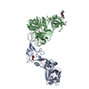
|
|---|---|
| 1 |
|
-Components
| #1: Protein | Mass: 37321.785 Da / Num. of mol.: 1 / Fragment: RBD Source method: isolated from a genetically manipulated source Source: (gene. exp.) Human coronavirus HKU1 (isolate N1) / Gene: S, 3 / Production host: Homo sapiens (human) / References: UniProt: Q5MQD0 | ||||
|---|---|---|---|---|---|
| #2: Protein | Mass: 46232.004 Da / Num. of mol.: 1 Source method: isolated from a genetically manipulated source Source: (gene. exp.) Homo sapiens (human) / Gene: TMPRSS2, PRSS10 / Production host: Homo sapiens (human)References: UniProt: O15393, transmembrane protease serine 2 | ||||
| #3: Polysaccharide | 2-acetamido-2-deoxy-beta-D-glucopyranose-(1-4)-2-acetamido-2-deoxy-beta-D-glucopyranose Source method: isolated from a genetically manipulated source | ||||
| #4: Sugar |   Type: D-saccharide, beta linking / Mass: 221.208 Da / Num. of mol.: 3 / Source method: obtained synthetically / Formula: C8H15NO6 Type: D-saccharide, beta linking / Mass: 221.208 Da / Num. of mol.: 3 / Source method: obtained synthetically / Formula: C8H15NO6Has ligand of interest | N | Has protein modification | Y | |
-Experimental details
-Experiment
| Experiment | Method: ELECTRON MICROSCOPY |
|---|---|
| EM experiment | Aggregation state: PARTICLE / 3D reconstruction method: single particle reconstruction |
- Sample preparation
| Component | Name: HKU1 RBD bound to the human TMPRSS2 receptor / Type: COMPLEX / Entity ID: #1-#2 / Source: RECOMBINANT |
|---|---|
| Molecular weight | Experimental value: NO |
| Source (natural) | Organism: Homo sapiens (human) |
| Source (recombinant) | Organism: Homo sapiens (human) |
| Buffer solution | pH: 8 |
| Specimen | Embedding applied: NO / Shadowing applied: NO / Staining applied: NO / Vitrification applied: YES |
| Vitrification | Cryogen name: ETHANE |
- Electron microscopy imaging
| Experimental equipment |  Model: Titan Krios / Image courtesy: FEI Company |
|---|---|
| Microscopy | Model: FEI TITAN KRIOS |
| Electron gun | Electron source:  FIELD EMISSION GUN / Accelerating voltage: 300 kV / Illumination mode: FLOOD BEAM FIELD EMISSION GUN / Accelerating voltage: 300 kV / Illumination mode: FLOOD BEAM |
| Electron lens | Mode: BRIGHT FIELD / Nominal defocus max: 3500 nm / Nominal defocus min: 500 nm |
| Image recording | Electron dose: 60 e/Å2 / Film or detector model: GATAN K3 (6k x 4k) |
- Processing
| EM software |
| |||||||||
|---|---|---|---|---|---|---|---|---|---|---|
| CTF correction | Type: PHASE FLIPPING AND AMPLITUDE CORRECTION | |||||||||
| 3D reconstruction | Resolution: 2.9 Å / Resolution method: FSC 0.143 CUT-OFF / Num. of particles: 810357 / Symmetry type: POINT |
