National Institutes of Health/National Institute of General Medical Sciences (NIH/NIGMS)
R35-GM142797
United States
Citation
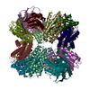
Journal: Int J Mol Sci / Year: 2024 Title: Mitigating the Blurring Effect of CryoEM Averaging on a Flexible and Highly Symmetric Protein Complex through Sub-Particle Reconstruction. Authors: Diana S Suder / Shane Gonen / Abstract: Many macromolecules are inherently flexible as a feature of their structure and function. During single-particle CryoEM processing, flexible protein regions can be detrimental to high-resolution ...Many macromolecules are inherently flexible as a feature of their structure and function. During single-particle CryoEM processing, flexible protein regions can be detrimental to high-resolution reconstruction as signals from thousands of particles are averaged together. This "blurring" effect can be difficult to overcome and is possibly more pronounced when averaging highly symmetric complexes. Approaches to mitigating flexibility during CryoEM processing are becoming increasingly critical as the technique advances and is applied to more dynamic proteins and complexes. Here, we detail the use of sub-particle averaging and signal subtraction techniques to precisely target and resolve flexible DARPin protein attachments on a designed tetrahedrally symmetric protein scaffold called DARP14. Particles are first aligned as full complexes, and then the symmetry is reduced by alignment and focused refinement of the constituent subunits. The final reconstructions we obtained were vastly improved over the fully symmetric reconstructions, with observable secondary structure and side-chain placement. Additionally, we were also able to reconstruct the core region of the scaffold to 2.7 Å. The data processing protocol outlined here is applicable to other dynamic and symmetric protein complexes, and our improved maps could allow for new structure-guided variant designs of DARP14.
History
Deposition
Dec 18, 2023
Deposition site: RCSB / Processing site: RCSB
Revision 1.0
Jun 5, 2024
Provider: repository / Type: Initial release
Revision 1.0
Jun 5, 2024
Data content type: EM metadata / Data content type: EM metadata / Provider: repository / Type: Initial release
Revision 1.0
Jun 5, 2024
Data content type: FSC / Data content type: FSC / Provider: repository / Type: Initial release
Revision 1.0
Jun 5, 2024
Data content type: Half map / Part number: 1 / Data content type: Half map / Provider: repository / Type: Initial release
Revision 1.0
Jun 5, 2024
Data content type: Half map / Part number: 2 / Data content type: Half map / Provider: repository / Type: Initial release
Revision 1.0
Jun 5, 2024
Data content type: Image / Data content type: Image / Provider: repository / Type: Initial release
Revision 1.0
Jun 5, 2024
Data content type: Primary map / Data content type: Primary map / Provider: repository / Type: Initial release
Revision 1.0
Jun 5, 2024
Data content type: FSC / Data content type: FSC / Provider: repository / Type: Initial release
Revision 1.0
Jun 5, 2024
Data content type: Half map / Part number: 1 / Data content type: Half map / Provider: repository / Type: Initial release
Revision 1.0
Jun 5, 2024
Data content type: Half map / Part number: 2 / Data content type: Half map / Provider: repository / Type: Initial release
Revision 1.0
Jun 5, 2024
Data content type: Image / Data content type: Image / Provider: repository / Type: Initial release
Revision 1.0
Jun 5, 2024
Data content type: Primary map / Data content type: Primary map / Provider: repository / Type: Initial release
Data content type: EM metadata / Data content type: EM metadata / EM metadata / Group: Data processing / Experimental summary / Data content type: EM metadata / EM metadata / Category: em_admin / em_software / Data content type: EM metadata / EM metadata / Item: _em_admin.last_update / _em_software.name
Mass: 32208.723 Da / Num. of mol.: 1 Source method: isolated from a genetically manipulated source Details: with a helical connection to a chain of a designed protein scaffold Source: (gene. exp.) synthetic construct (others) / Production host: Escherichia coli BL21(DE3) (bacteria)
Has protein modification
N
-
Experimental details
-
Experiment
Experiment
Method: ELECTRON MICROSCOPY
EM experiment
Aggregation state: PARTICLE / 3D reconstruction method: single particle reconstruction
-
Sample preparation
Component
Name: DARPin with a helical connection to a chain of a designed protein scaffold Type: COMPLEX / Entity ID: all / Source: RECOMBINANT
Molecular weight
Experimental value: NO
Source (natural)
Organism: synthetic construct (others)
Source (recombinant)
Organism: Escherichia coli BL21(DE3) (bacteria)
Buffer solution
pH: 8
Specimen
Embedding applied: NO / Shadowing applied: NO / Staining applied: NO / Vitrification applied: YES
Vitrification
Cryogen name: ETHANE
-
Electron microscopy imaging
Experimental equipment
Model: Titan Krios / Image courtesy: FEI Company
Microscopy
Model: FEI TITAN KRIOS
Electron gun
Electron source: FIELD EMISSION GUN / Accelerating voltage: 300 kV / Illumination mode: OTHER
Electron dose: 30 e/Å2 / Film or detector model: GATAN K2 SUMMIT (4k x 4k)
-
Processing
EM software
Name: PHENIX / Category: model refinement
CTF correction
Type: PHASE FLIPPING AND AMPLITUDE CORRECTION
Symmetry
Point symmetry: C1 (asymmetric)
3D reconstruction
Resolution: 4 Å / Resolution method: FSC 0.143 CUT-OFF / Num. of particles: 287323 Details: resolution ranges drastically across the map, though. Symmetry type: POINT
Refine LS restraints
Refine-ID
Type
Dev ideal
Number
ELECTRONMICROSCOPY
f_bond_d
0.003
4489
ELECTRONMICROSCOPY
f_angle_d
0.503
6049
ELECTRONMICROSCOPY
f_dihedral_angle_d
3.699
606
ELECTRONMICROSCOPY
f_chiral_restr
0.037
715
ELECTRONMICROSCOPY
f_plane_restr
0.003
768
+
About Yorodumi
-
News
-
Feb 9, 2022. New format data for meta-information of EMDB entries
New format data for meta-information of EMDB entries
Version 3 of the EMDB header file is now the official format.
The previous official version 1.9 will be removed from the archive.
In the structure databanks used in Yorodumi, some data are registered as the other names, "COVID-19 virus" and "2019-nCoV". Here are the details of the virus and the list of structure data.
Jan 31, 2019. EMDB accession codes are about to change! (news from PDBe EMDB page)
EMDB accession codes are about to change! (news from PDBe EMDB page)
The allocation of 4 digits for EMDB accession codes will soon come to an end. Whilst these codes will remain in use, new EMDB accession codes will include an additional digit and will expand incrementally as the available range of codes is exhausted. The current 4-digit format prefixed with “EMD-” (i.e. EMD-XXXX) will advance to a 5-digit format (i.e. EMD-XXXXX), and so on. It is currently estimated that the 4-digit codes will be depleted around Spring 2019, at which point the 5-digit format will come into force.
The EM Navigator/Yorodumi systems omit the EMD- prefix.
Related info.:Q: What is EMD? / ID/Accession-code notation in Yorodumi/EM Navigator
Yorodumi is a browser for structure data from EMDB, PDB, SASBDB, etc.
This page is also the successor to EM Navigator detail page, and also detail information page/front-end page for Omokage search.
The word "yorodu" (or yorozu) is an old Japanese word meaning "ten thousand". "mi" (miru) is to see.
Related info.:EMDB / PDB / SASBDB / Comparison of 3 databanks / Yorodumi Search / Aug 31, 2016. New EM Navigator & Yorodumi / Yorodumi Papers / Jmol/JSmol / Function and homology information / Changes in new EM Navigator and Yorodumi
 Movie
Movie Controller


 Open data
 Basic information
 Components
 Keywords
 Authors
 United States, 1items
  Citation
 Structure visualization
 Molmil
 Downloads & links
 Other downloads


 PDBj
PDBj
 Assembly

 Sample preparation
 Electron microscopy imaging
 FIELD EMISSION GUN / Accelerating voltage: 300 kV / Illumination mode: OTHER
 Processing