[English] 日本語
Yorodumi
- PDB-8vdz: A designed tetrahedral protein scaffold - DARP14 -

+
Open data


ID or keywords:

Loading...

-
Basic information

Entry
Database: PDB / ID: 8vdz
TitleA designed tetrahedral protein scaffold - DARP14
Components
  • Subunit A
  • Subunit B
KeywordsDE NOVO PROTEIN / scaffold / tetrahedral / darpin / symmetrical
Biological speciessynthetic construct (others)
MethodELECTRON MICROSCOPY / single particle reconstruction / cryo EM / Resolution: 2.7 Å
AuthorsSuder, D.S. / Gonen, S.
Funding support United States, 1items
OrganizationGrant numberCountry
National Institutes of Health/National Institute of General Medical Sciences (NIH/NIGMS)R35-GM142797 United States
Citation
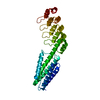
Journal: Int J Mol Sci / Year: 2024
Title: Mitigating the Blurring Effect of CryoEM Averaging on a Flexible and Highly Symmetric Protein Complex through Sub-Particle Reconstruction.
Authors: Diana S Suder / Shane Gonen /
Abstract: Many macromolecules are inherently flexible as a feature of their structure and function. During single-particle CryoEM processing, flexible protein regions can be detrimental to high-resolution ...Many macromolecules are inherently flexible as a feature of their structure and function. During single-particle CryoEM processing, flexible protein regions can be detrimental to high-resolution reconstruction as signals from thousands of particles are averaged together. This "blurring" effect can be difficult to overcome and is possibly more pronounced when averaging highly symmetric complexes. Approaches to mitigating flexibility during CryoEM processing are becoming increasingly critical as the technique advances and is applied to more dynamic proteins and complexes. Here, we detail the use of sub-particle averaging and signal subtraction techniques to precisely target and resolve flexible DARPin protein attachments on a designed tetrahedrally symmetric protein scaffold called DARP14. Particles are first aligned as full complexes, and then the symmetry is reduced by alignment and focused refinement of the constituent subunits. The final reconstructions we obtained were vastly improved over the fully symmetric reconstructions, with observable secondary structure and side-chain placement. Additionally, we were also able to reconstruct the core region of the scaffold to 2.7 Å. The data processing protocol outlined here is applicable to other dynamic and symmetric protein complexes, and our improved maps could allow for new structure-guided variant designs of DARP14.
History
DepositionDec 18, 2023Deposition site: RCSB / Processing site: RCSB
Revision 1.0Jun 5, 2024Provider: repository / Type: Initial release
Revision 1.1Jul 3, 2024Group: Data collection / Database references / Category: citation / em_admin
Item: _citation.pdbx_database_id_PubMed / _citation.title / _em_admin.last_update

-
Structure visualization

Structure viewerMolecule:
MolmilJmol/JSmol

Downloads & links

-
Assembly

Deposited unit
A: Subunit A
B: Subunit A
C: Subunit A
D: Subunit A
E: Subunit B
F: Subunit B
G: Subunit B
H: Subunit B
I: Subunit A
J: Subunit A
K: Subunit A
L: Subunit A
M: Subunit B
N: Subunit B
O: Subunit B
P: Subunit B
Q: Subunit A
R: Subunit A
S: Subunit A
T: Subunit A
U: Subunit B
V: Subunit B
W: Subunit B
X: Subunit B


Theoretical massNumber of molelcules
Total (without water)340,86924
Polymers340,86924
Non-polymers00
Water00
1


  • Idetical with deposited unit
  • defined by author
  • Evidence: electron microscopy
TypeNameSymmetry operationNumber
identity operation1_5551

-
Components

#1: Protein
Subunit A


Mass: 15775.438 Da / Num. of mol.: 12
Source method: isolated from a genetically manipulated source
Source: (gene. exp.) synthetic construct (others) / Production host: Escherichia coli BL21(DE3) (bacteria) / References: corrinoid adenosyltransferase
#2: Protein
Subunit B


Mass: 12630.332 Da / Num. of mol.: 12
Source method: isolated from a genetically manipulated source
Source: (gene. exp.) synthetic construct (others) / Production host: Escherichia coli BL21(DE3) (bacteria)

-
Experimental details

-
Experiment

ExperimentMethod: ELECTRON MICROSCOPY
EM experimentAggregation state: PARTICLE / 3D reconstruction method: single particle reconstruction

-
Sample preparation

Component
IDNameTypeEntity IDParent-IDSource
1A12B12 tetrahedral complex of a designed protein scaffold called DARP14COMPLEXall0RECOMBINANT
2Subunit A in a designed protein scaffold called DARP14COMPLEX#11RECOMBINANT
3Subunit B in a designed protein scaffold called DARP14COMPLEX#21RECOMBINANT
Molecular weight
IDEntity assembly-IDExperimental value
11NO
21NO
33
Source (natural)
IDEntity assembly-IDOrganismNcbi tax-ID
21synthetic construct (others)32630
32synthetic construct (others)32630
43synthetic construct (others)32630
Source (recombinant)
IDEntity assembly-IDOrganismNcbi tax-ID
21Escherichia coli BL21(DE3) (bacteria)469008
32Escherichia coli BL21(DE3) (bacteria)469008
43Escherichia coli BL21(DE3) (bacteria)469008
Buffer solutionpH: 8
SpecimenConc.: 0.5 mg/ml / Embedding applied: NO / Shadowing applied: NO / Staining applied: NO / Vitrification applied: YES
Specimen supportGrid material: COPPER / Grid mesh size: 200 divisions/in.
VitrificationCryogen name: ETHANE

-
Electron microscopy imaging

Experimental equipment
Model: Titan Krios / Image courtesy: FEI Company
MicroscopyModel: FEI TITAN KRIOS
Electron gunElectron source: FIELD EMISSION GUN / Accelerating voltage: 300 kV / Illumination mode: OTHER
Electron lensMode: BRIGHT FIELD / Nominal defocus max: 2500 nm / Nominal defocus min: 700 nm
Image recordingElectron dose: 30 e/Å2 / Film or detector model: GATAN K2 SUMMIT (4k x 4k)

-
Processing

EM software
IDNameCategory
7UCSF ChimeraXmodel fitting
9RELIONinitial Euler assignment
10RELIONfinal Euler assignment
11RELIONclassification
12RELION3D reconstruction
13PHENIXmodel refinement
CTF correctionType: PHASE FLIPPING AND AMPLITUDE CORRECTION
SymmetryPoint symmetry: T (tetrahedral)
3D reconstructionResolution: 2.7 Å / Resolution method: FSC 0.143 CUT-OFF / Num. of particles: 277091 / Symmetry type: POINT
Atomic model buildingSpace: REAL / Details: Phenix real space refine
Refine LS restraints
Refine-IDTypeDev idealNumber
ELECTRON MICROSCOPYf_bond_d0.00323936
ELECTRON MICROSCOPYf_angle_d0.42232223
ELECTRON MICROSCOPYf_dihedral_angle_d3.5233339
ELECTRON MICROSCOPYf_chiral_restr0.0383852
ELECTRON MICROSCOPYf_plane_restr0.0034091

+
About Yorodumi

-
News

-
Feb 9, 2022. New format data for meta-information of EMDB entries

New format data for meta-information of EMDB entries

  • Version 3 of the EMDB header file is now the official format.
  • The previous official version 1.9 will be removed from the archive.

Related info.:EMDB header

External links:wwPDB to switch to version 3 of the EMDB data model

-
Aug 12, 2020. Covid-19 info

Covid-19 info

URL: https://pdbjlc1.pdbj.org/emnavi/covid19.php

New page: Covid-19 featured information page in EM Navigator.

Related info.:Covid-19 info / Mar 5, 2020. Novel coronavirus structure data

+
Mar 5, 2020. Novel coronavirus structure data

Novel coronavirus structure data

Related info.:Yorodumi Speices / Aug 12, 2020. Covid-19 info

External links:COVID-19 featured content - PDBj / Molecule of the Month (242):Coronavirus Proteases

+
Jan 31, 2019. EMDB accession codes are about to change! (news from PDBe EMDB page)

EMDB accession codes are about to change! (news from PDBe EMDB page)

  • The allocation of 4 digits for EMDB accession codes will soon come to an end. Whilst these codes will remain in use, new EMDB accession codes will include an additional digit and will expand incrementally as the available range of codes is exhausted. The current 4-digit format prefixed with “EMD-” (i.e. EMD-XXXX) will advance to a 5-digit format (i.e. EMD-XXXXX), and so on. It is currently estimated that the 4-digit codes will be depleted around Spring 2019, at which point the 5-digit format will come into force.
  • The EM Navigator/Yorodumi systems omit the EMD- prefix.

Related info.:Q: What is EMD? / ID/Accession-code notation in Yorodumi/EM Navigator

External links:EMDB Accession Codes are Changing Soon! / Contact to PDBj

+
Jul 12, 2017. Major update of PDB

Major update of PDB

  • wwPDB released updated PDB data conforming to the new PDBx/mmCIF dictionary.
  • This is a major update changing the version number from 4 to 5, and with Remediation, in which all the entries are updated.
  • In this update, many items about electron microscopy experimental information are reorganized (e.g. em_software).
  • Now, EM Navigator and Yorodumi are based on the updated data.

External links:wwPDB Remediation / Enriched Model Files Conforming to OneDep Data Standards Now Available in the PDB FTP Archive

-
Yorodumi

Thousand views of thousand structures

  • Yorodumi is a browser for structure data from EMDB, PDB, SASBDB, etc.
  • This page is also the successor to EM Navigator detail page, and also detail information page/front-end page for Omokage search.
  • The word "yorodu" (or yorozu) is an old Japanese word meaning "ten thousand". "mi" (miru) is to see.

Related info.:EMDB / PDB / SASBDB / Comparison of 3 databanks / Yorodumi Search / Aug 31, 2016. New EM Navigator & Yorodumi / Yorodumi Papers / Jmol/JSmol / Function and homology information / Changes in new EM Navigator and Yorodumi

Read more