+ Open data
- Basic information
| Entry | Database: PDB / ID: 8v45 | ||||||
|---|---|---|---|---|---|---|---|
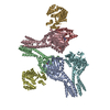
| Title | CryoEM structure of AriA-Ocr complex | ||||||
 Components Components |
| ||||||
 Keywords Keywords | IMMUNE SYSTEM / Bacterial Defense system / Toxin-antitoxin system / AriAB / PARIS | ||||||
| Function / homology | B-form DNA mimic Ocr / DNA mimic ocr / Protein Ocr / symbiont-mediated evasion of host restriction-modification system / symbiont-mediated suppression of host innate immune response / ADENOSINE-5'-TRIPHOSPHATE / : / Protein Ocr Function and homology information Function and homology information | ||||||
| Biological species |    Escherichia phage T7 (virus) Escherichia phage T7 (virus) | ||||||
| Method | ELECTRON MICROSCOPY / single particle reconstruction / cryo EM / Resolution: 3.63 Å | ||||||
 Authors Authors | Deep, A. / Corbett, K.D. | ||||||
| Funding support |  United States, 1items United States, 1items
| ||||||
 Citation Citation |  Journal: Nature / Year: 2024 Journal: Nature / Year: 2024Title: Architecture and activation mechanism of the bacterial PARIS defence system. Authors: Amar Deep / Qishan Liang / Eray Enustun / Joe Pogliano / Kevin D Corbett /  Abstract: Bacteria and their viruses (bacteriophages or phages) are engaged in an intense evolutionary arms race. While the mechanisms of many bacterial antiphage defence systems are known, how these systems ...Bacteria and their viruses (bacteriophages or phages) are engaged in an intense evolutionary arms race. While the mechanisms of many bacterial antiphage defence systems are known, how these systems avoid toxicity outside infection yet activate quickly after infection is less well understood. Here we show that the bacterial phage anti-restriction-induced system (PARIS) operates as a toxin-antitoxin system, in which the antitoxin AriA sequesters and inactivates the toxin AriB until triggered by the T7 phage counterdefence protein Ocr. Using cryo-electron microscopy, we show that AriA is related to SMC-family ATPases but assembles into a distinctive homohexameric complex through two oligomerization interfaces. In uninfected cells, the AriA hexamer binds to up to three monomers of AriB, maintaining them in an inactive state. After Ocr binding, the AriA hexamer undergoes a structural rearrangement, releasing AriB and allowing it to dimerize and activate. AriB is a toprim/OLD-family nuclease, the activation of which arrests cell growth and inhibits phage propagation by globally inhibiting protein translation through specific cleavage of a lysine tRNA. Collectively, our findings reveal the intricate molecular mechanisms of a bacterial defence system triggered by a phage counterdefence protein, and highlight how an SMC-family ATPase has been adapted as a bacterial infection sensor. | ||||||
| History |
|
- Structure visualization
| Structure viewer | Molecule:  Molmil Molmil Jmol/JSmol Jmol/JSmol |
|---|
- Downloads & links
- Download
| PDBx/mmCIF format |  8v45.cif.gz 8v45.cif.gz | 438.6 KB | Display |  PDBx/mmCIF format PDBx/mmCIF format |
|---|---|---|---|---|
| PDB format |  pdb8v45.ent.gz pdb8v45.ent.gz | 359.8 KB | Display |  PDB format PDB format |
| PDBx/mmJSON format |  8v45.json.gz 8v45.json.gz | Tree view |  PDBx/mmJSON format PDBx/mmJSON format | |
| Others |  Other downloads Other downloads |
-Validation report
| Summary document |  8v45_validation.pdf.gz 8v45_validation.pdf.gz | 1.6 MB | Display |  wwPDB validaton report wwPDB validaton report |
|---|---|---|---|---|
| Full document |  8v45_full_validation.pdf.gz 8v45_full_validation.pdf.gz | 1.6 MB | Display | |
| Data in XML |  8v45_validation.xml.gz 8v45_validation.xml.gz | 75.5 KB | Display | |
| Data in CIF |  8v45_validation.cif.gz 8v45_validation.cif.gz | 114.8 KB | Display | |
| Arichive directory |  https://data.pdbj.org/pub/pdb/validation_reports/v4/8v45 https://data.pdbj.org/pub/pdb/validation_reports/v4/8v45 ftp://data.pdbj.org/pub/pdb/validation_reports/v4/8v45 ftp://data.pdbj.org/pub/pdb/validation_reports/v4/8v45 | HTTPS FTP |
-Related structure data
| Related structure data |  42965MC  8v46C  8v47C  8v48C  8v49C M: map data used to model this data C: citing same article ( |
|---|---|
| Similar structure data | Similarity search - Function & homology  F&H Search F&H Search |
- Links
Links
- Assembly
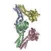
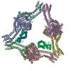
| Deposited unit | 
|
|---|---|
| 1 |
|
- Components
| #1: Protein | Mass: 53288.098 Da / Num. of mol.: 6 Source method: isolated from a genetically manipulated source Source: (gene. exp.)   #2: Protein | Mass: 13819.015 Da / Num. of mol.: 2 Source method: isolated from a genetically manipulated source Source: (gene. exp.)   Escherichia phage T7 (virus) / Gene: 0.3 / Production host: Escherichia phage T7 (virus) / Gene: 0.3 / Production host:  #3: Chemical | ChemComp-ATP / Has ligand of interest | Y | Has protein modification | N | |
|---|
-Experimental details
-Experiment
| Experiment | Method: ELECTRON MICROSCOPY |
|---|---|
| EM experiment | Aggregation state: PARTICLE / 3D reconstruction method: single particle reconstruction |
- Sample preparation
| Component | Name: AriA hexamer with two monomers of Ocr / Type: COMPLEX / Details: AriA(6)-Ocr(2) / Entity ID: #1-#2 / Source: RECOMBINANT | ||||||||||||||||||||||||||||||
|---|---|---|---|---|---|---|---|---|---|---|---|---|---|---|---|---|---|---|---|---|---|---|---|---|---|---|---|---|---|---|---|
| Molecular weight | Experimental value: NO | ||||||||||||||||||||||||||||||
| Source (natural) | Organism:  | ||||||||||||||||||||||||||||||
| Source (recombinant) | Organism:  | ||||||||||||||||||||||||||||||
| Buffer solution | pH: 7.4 | ||||||||||||||||||||||||||||||
| Buffer component |
| ||||||||||||||||||||||||||||||
| Specimen | Conc.: 2 mg/ml / Embedding applied: NO / Shadowing applied: NO / Staining applied: NO / Vitrification applied: YES / Details: Sample was a mixture of three proteins. | ||||||||||||||||||||||||||||||
| Specimen support | Grid material: GOLD / Grid mesh size: 300 divisions/in. / Grid type: UltrAuFoil R1.2/1.3 | ||||||||||||||||||||||||||||||
| Vitrification | Instrument: FEI VITROBOT MARK IV / Cryogen name: ETHANE / Humidity: 95 % / Chamber temperature: 278 K |
- Electron microscopy imaging
| Experimental equipment |  Model: Titan Krios / Image courtesy: FEI Company |
|---|---|
| Microscopy | Model: FEI TITAN KRIOS |
| Electron gun | Electron source:  FIELD EMISSION GUN / Accelerating voltage: 300 kV / Illumination mode: FLOOD BEAM FIELD EMISSION GUN / Accelerating voltage: 300 kV / Illumination mode: FLOOD BEAM |
| Electron lens | Mode: BRIGHT FIELD / Nominal defocus max: 2000 nm / Nominal defocus min: 500 nm / Cs: 2.7 mm |
| Image recording | Electron dose: 50 e/Å2 / Film or detector model: FEI FALCON IV (4k x 4k) / Num. of grids imaged: 1 / Num. of real images: 6821 |
| EM imaging optics | Energyfilter name: TFS Selectris X / Energyfilter slit width: 20 eV |
- Processing
| EM software | Name: PHENIX / Version: 1.21rc1_5127 / Category: model refinement | ||||||||||||||||||||||||
|---|---|---|---|---|---|---|---|---|---|---|---|---|---|---|---|---|---|---|---|---|---|---|---|---|---|
| CTF correction | Type: NONE | ||||||||||||||||||||||||
| 3D reconstruction | Resolution: 3.63 Å / Resolution method: FSC 0.143 CUT-OFF / Num. of particles: 99697 / Algorithm: FOURIER SPACE / Symmetry type: POINT | ||||||||||||||||||||||||
| Atomic model building | Protocol: AB INITIO MODEL / Space: REAL | ||||||||||||||||||||||||
| Refinement | Cross valid method: NONE Stereochemistry target values: GeoStd + Monomer Library + CDL v1.2 | ||||||||||||||||||||||||
| Displacement parameters | Biso mean: 169.47 Å2 | ||||||||||||||||||||||||
| Refine LS restraints |
|
 Movie
Movie Controller


 PDBj
PDBj

