[English] 日本語
 Yorodumi
- PDB-8v1s: Herpes simplex virus 1 polymerase holoenzyme bound to mismatched ... -
+ Open data
- Basic information
| Entry | Database: PDB / ID: 8v1s | |||||||||
|---|---|---|---|---|---|---|---|---|---|---|
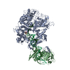
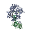
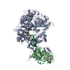
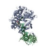
| Title | Herpes simplex virus 1 polymerase holoenzyme bound to mismatched DNA in editing conformation | |||||||||
 Components Components |
| |||||||||
 Keywords Keywords | TRANSFERASE/DNA / herpes simplex virus / replication / DNA polymerase holoenzyme / editing conformation / TRANSFERASE-DNA complex | |||||||||
| Function / homology |  Function and homology information Function and homology informationbidirectional double-stranded viral DNA replication / exonuclease activity / DNA-templated DNA replication / RNA-DNA hybrid ribonuclease activity / DNA-directed DNA polymerase / DNA-directed DNA polymerase activity / DNA replication / nucleotide binding / host cell nucleus / DNA binding Similarity search - Function | |||||||||
| Biological species |   Human alphaherpesvirus 1 strain KOS Human alphaherpesvirus 1 strain KOSsynthetic construct (others) | |||||||||
| Method | ELECTRON MICROSCOPY / single particle reconstruction / cryo EM / Resolution: 3.26 Å | |||||||||
 Authors Authors | Pan, J. / Abraham, J. / Coen, D.M. / Shankar, S. / Yang, P. / Hogle, J. | |||||||||
| Funding support |  United States, 2items United States, 2items
| |||||||||
 Citation Citation |  Journal: Cell / Year: 2024 Journal: Cell / Year: 2024Title: Viral DNA polymerase structures reveal mechanisms of antiviral drug resistance. Authors: Sundaresh Shankar / Junhua Pan / Pan Yang / Yuemin Bian / Gábor Oroszlán / Zishuo Yu / Purba Mukherjee / David J Filman / James M Hogle / Mrinal Shekhar / Donald M Coen / Jonathan Abraham /    Abstract: DNA polymerases are important drug targets, and many structural studies have captured them in distinct conformations. However, a detailed understanding of the impact of polymerase conformational ...DNA polymerases are important drug targets, and many structural studies have captured them in distinct conformations. However, a detailed understanding of the impact of polymerase conformational dynamics on drug resistance is lacking. We determined cryoelectron microscopy (cryo-EM) structures of DNA-bound herpes simplex virus polymerase holoenzyme in multiple conformations and interacting with antivirals in clinical use. These structures reveal how the catalytic subunit Pol and the processivity factor UL42 bind DNA to promote processive DNA synthesis. Unexpectedly, in the absence of an incoming nucleotide, we observed Pol in multiple conformations with the closed state sampled by the fingers domain. Drug-bound structures reveal how antivirals may selectively bind enzymes that more readily adopt the closed conformation. Molecular dynamics simulations and the cryo-EM structure of a drug-resistant mutant indicate that some resistance mutations modulate conformational dynamics rather than directly impacting drug binding, thus clarifying mechanisms that drive drug selectivity. | |||||||||
| History |
|
- Structure visualization
| Structure viewer | Molecule:  Molmil Molmil Jmol/JSmol Jmol/JSmol |
|---|
- Downloads & links
- Download
| PDBx/mmCIF format |  8v1s.cif.gz 8v1s.cif.gz | 319 KB | Display |  PDBx/mmCIF format PDBx/mmCIF format |
|---|---|---|---|---|
| PDB format |  pdb8v1s.ent.gz pdb8v1s.ent.gz | 242.2 KB | Display |  PDB format PDB format |
| PDBx/mmJSON format |  8v1s.json.gz 8v1s.json.gz | Tree view |  PDBx/mmJSON format PDBx/mmJSON format | |
| Others |  Other downloads Other downloads |
-Validation report
| Summary document |  8v1s_validation.pdf.gz 8v1s_validation.pdf.gz | 1.5 MB | Display |  wwPDB validaton report wwPDB validaton report |
|---|---|---|---|---|
| Full document |  8v1s_full_validation.pdf.gz 8v1s_full_validation.pdf.gz | 1.5 MB | Display | |
| Data in XML |  8v1s_validation.xml.gz 8v1s_validation.xml.gz | 56 KB | Display | |
| Data in CIF |  8v1s_validation.cif.gz 8v1s_validation.cif.gz | 84.3 KB | Display | |
| Arichive directory |  https://data.pdbj.org/pub/pdb/validation_reports/v1/8v1s https://data.pdbj.org/pub/pdb/validation_reports/v1/8v1s ftp://data.pdbj.org/pub/pdb/validation_reports/v1/8v1s ftp://data.pdbj.org/pub/pdb/validation_reports/v1/8v1s | HTTPS FTP |
-Related structure data
| Related structure data |  42889MC  8exxC  8v1qC  8v1rC  8v1tC C: citing same article ( M: map data used to model this data |
|---|---|
| Similar structure data | Similarity search - Function & homology  F&H Search F&H Search |
- Links
Links
- Assembly
| Deposited unit | 
|
|---|---|
| 1 |
|
- Components
| #1: Protein | Mass: 133614.344 Da / Num. of mol.: 1 Source method: isolated from a genetically manipulated source Details: Herpes simplex virus type 1 (KOS strain) DNA polymerase catalytic subunit UL30 with its N-terminal 42 residues deleted and replaced by an N-terminal poly-histidine tag in the expression construct Source: (gene. exp.)   Human alphaherpesvirus 1 strain KOS / Gene: UL30, HHV1gp046 / Cell line (production host): Sf9 / Production host: Human alphaherpesvirus 1 strain KOS / Gene: UL30, HHV1gp046 / Cell line (production host): Sf9 / Production host:  |
|---|---|
| #2: Protein | Mass: 36346.180 Da / Num. of mol.: 1 Source method: isolated from a genetically manipulated source Details: Herpes simplex virus 1 (KOS strain) DNA polymerase processivity factor UL42 residues 1-340 tagged with a prescission protease cleavage site followed by a maltose binding proten (MBP) tag in ...Details: Herpes simplex virus 1 (KOS strain) DNA polymerase processivity factor UL42 residues 1-340 tagged with a prescission protease cleavage site followed by a maltose binding proten (MBP) tag in the expression construct Source: (gene. exp.)   Human alphaherpesvirus 1 strain KOS / Gene: UL42, HHV1gp061 / Plasmid: PMAL-2C / Production host: Human alphaherpesvirus 1 strain KOS / Gene: UL42, HHV1gp061 / Plasmid: PMAL-2C / Production host:  |
| #3: DNA chain | Mass: 10227.623 Da / Num. of mol.: 1 / Source method: obtained synthetically Details: Contains phosphorothioated 3' mismatch with template DNA Source: (synth.) synthetic construct (others) |
| #4: DNA chain | Mass: 15223.799 Da / Num. of mol.: 1 / Source method: obtained synthetically / Source: (synth.) synthetic construct (others) |
| #5: Chemical | ChemComp-MG / |
| Has ligand of interest | N |
| Has protein modification | N |
-Experimental details
-Experiment
| Experiment | Method: ELECTRON MICROSCOPY |
|---|---|
| EM experiment | Aggregation state: PARTICLE / 3D reconstruction method: single particle reconstruction |
- Sample preparation
| Component | Name: HSV-1 DNA polymerase holoenzyme editing complex / Type: COMPLEX Details: Herpes simplex virus type 1 polymerase holoenzyme UL30:UL42 in complex with primer DNA that has a 3'-end mismatch with template DNA Entity ID: #1-#4 / Source: RECOMBINANT |
|---|---|
| Molecular weight | Value: 0.197 MDa / Experimental value: NO |
| Source (natural) | Organism:   Human herpesvirus 1 (strain KOS) Human herpesvirus 1 (strain KOS) |
| Source (recombinant) | Organism:  |
| Buffer solution | pH: 7.5 Details: 25 mM HEPES, pH 7.5, 150 mM NaCl, 2 mM tris(2-carboxyethyl)phosphine (TCEP) |
| Specimen | Conc.: 1.38 mg/ml / Embedding applied: NO / Shadowing applied: NO / Staining applied: NO / Vitrification applied: YES / Details: This sample was monodisperse. |
| Specimen support | Details: GRIDS WERE GLOW DISCHARGED IN A PELCO EASIGLOW AT 15 MA FOR 30 SECONDS UNDER 0.39 MBAR PRESSURE. Grid material: COPPER / Grid mesh size: 400 divisions/in. / Grid type: Quantifoil R1.2/1.3 |
| Vitrification | Instrument: FEI VITROBOT MARK IV / Cryogen name: ETHANE / Humidity: 100 % / Chamber temperature: 298 K Details: 3 microliters of sample were blotted for 3 seconds with filter paper saturated under 100% humidity prior to plunging. |
- Electron microscopy imaging
| Experimental equipment |  Model: Titan Krios / Image courtesy: FEI Company |
|---|---|
| Microscopy | Model: FEI TITAN KRIOS |
| Electron gun | Electron source:  FIELD EMISSION GUN / Accelerating voltage: 300 kV / Illumination mode: FLOOD BEAM FIELD EMISSION GUN / Accelerating voltage: 300 kV / Illumination mode: FLOOD BEAM |
| Electron lens | Mode: BRIGHT FIELD / Nominal magnification: 105000 X / Calibrated magnification: 60606 X / Nominal defocus max: 2500 nm / Nominal defocus min: 1000 nm / Calibrated defocus min: 1000 nm / Calibrated defocus max: 2500 nm / Cs: 2.7 mm / C2 aperture diameter: 50 µm / Alignment procedure: COMA FREE |
| Specimen holder | Cryogen: NITROGEN / Specimen holder model: FEI TITAN KRIOS AUTOGRID HOLDER / Temperature (max): 77 K / Temperature (min): 70 K |
| Image recording | Average exposure time: 1.5 sec. / Electron dose: 58.4 e/Å2 / Film or detector model: GATAN K3 (6k x 4k) / Num. of grids imaged: 1 / Num. of real images: 5076 |
| EM imaging optics | Energyfilter name: GIF Bioquantum / Energyfilter slit width: 20 eV |
| Image scans | Sampling size: 5 µm / Width: 5760 / Height: 4092 |
- Processing
| EM software |
| ||||||||||||||||||||||||||||||||||||||||||||
|---|---|---|---|---|---|---|---|---|---|---|---|---|---|---|---|---|---|---|---|---|---|---|---|---|---|---|---|---|---|---|---|---|---|---|---|---|---|---|---|---|---|---|---|---|---|
| CTF correction | Type: NONE | ||||||||||||||||||||||||||||||||||||||||||||
| Particle selection | Num. of particles selected: 3638013 | ||||||||||||||||||||||||||||||||||||||||||||
| Symmetry | Point symmetry: C1 (asymmetric) | ||||||||||||||||||||||||||||||||||||||||||||
| 3D reconstruction | Resolution: 3.26 Å / Resolution method: FSC 0.143 CUT-OFF / Num. of particles: 191794 / Algorithm: FOURIER SPACE / Num. of class averages: 1 / Symmetry type: POINT | ||||||||||||||||||||||||||||||||||||||||||||
| Atomic model building | B value: 54.86 / Protocol: OTHER / Space: REAL / Target criteria: correlation coefficient Details: rigid body, minimization_global, local_grid_search, adp refinement | ||||||||||||||||||||||||||||||||||||||||||||
| Refinement | Highest resolution: 3.26 Å |
 Movie
Movie Controller


 PDBj
PDBj


