- PDB-8uux: Murine norovirus capsid protein in the presence of 1mM calcium -
+
データを開く
IDまたはキーワード:
読み込み中...
-
基本情報
登録情報
データベース: PDB / ID: 8uux
タイトル
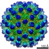
Murine norovirus capsid protein in the presence of 1mM calcium
要素
Capsid protein
キーワード
VIRAL PROTEIN / murine / norovirus / metals / activation
機能・相同性
Calicivirus coat protein C-terminal / Calicivirus coat protein C-terminal / Calicivirus coat protein / Calicivirus coat protein / Picornavirus/Calicivirus coat protein / Viral coat protein subunit / virus-mediated perturbation of host defense response / Capsid protein
National Institutes of Health/National Institute Of Allergy and Infectious Diseases (NIH/NIAID)
AI141465
米国
引用
ジャーナル: J Virol / 年: 2024 タイトル: The reversible activation of norovirus by metal ions. 著者: Michael Sherman / Faith Cox / Hong Smith / Mohamed H Habib / Stephanie Karst / Christiane E Wobus / Thomas J Smith / 要旨: Murine norovirus (MNV) undergoes extremely large conformational changes in response to the environment. The = 3 icosahedral capsid is composed of 180 copies of ~58-kDa VP1 comprised of N-terminus (N) ...Murine norovirus (MNV) undergoes extremely large conformational changes in response to the environment. The = 3 icosahedral capsid is composed of 180 copies of ~58-kDa VP1 comprised of N-terminus (N), shell (S), and C-terminal protruding (P) domains. At neutral pH, the P domains are loosely tethered to the shell and float ~15 Å above the surface. At low pH or in the presence of bile salts, the P domain drops onto the shell and this movement is accompanied by conformational changes within the P domain that enhance receptor interactions while blocking antibody binding. While previous crystallographic studies identified metal binding sites in the isolated P domain, the ~2.7-Å cryo-electron microscopy structures of MNV in the presence of Mg or Ca presented here show that metal ions can recapitulate the contraction observed at low pH or in the presence of bile. Further, we show that these conformational changes are reversed by dialysis against EDTA. As observed in the P domain crystal structures, metal ions bind to and contract the G'H' loop. This movement is correlated with the lifting of the C'D' loop and rotation of the P domain dimers about each other, exposing the bile salt binding pocket. Isothermal titration calorimetry experiments presented here demonstrate that the activation signals (bile salts, low pH, and metal ions) act in a synergistic manner that, individually, all result in the same activated structure. We present a model whereby these reversible conformational changes represent a uniquely dynamic and tissue-specific structural adaptation to the environment.IMPORTANCEThe highly mobile protruding domains on the calicivirus capsids are recognized by cell receptor(s) and antibodies. At neutral pH, they float ~15 Å above the shell but at low pH or in the presence of bile salts, they contract onto the surface. Concomitantly, changes within the P domain block antibody binding while enhancing receptor binding. While we previously demonstrated that metals also block antibody binding, it was unknown whether they might also cause similar conformational changes in the virion. Here, we present the near atomic cryo-electron microscopy structures of infectious murine norovirus (MNV) in the presence of calcium or magnesium ions. The metal ions reversibly induce the same P domain contraction as low pH and bile salts and act in a synergistic manner with the other stimuli. We propose that, unlike most other viruses, MNV facilely changes conformations as a unique means to escape immune surveillance as it moves through various tissues.
 ムービー
ムービー コントローラー


 データを開く
 基本情報
基本情報 要素
要素 キーワード
キーワード 機能・相同性情報
 Murine norovirus 1 (マウスノロウイルス 1)
 データ登録者
 米国, 1件
  引用
引用
 構造の表示
構造の表示 ダウンロードとリンク
 その他のダウンロード


 PDBj
PDBj
 集合体
集合体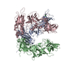

 分子量: 40.078 Da / 分子数: 3 / 由来タイプ: 合成 / 式: Ca / タイプ: SUBJECT OF INVESTIGATION
 分子量: 18.015 Da / 分子数: 310 / 由来タイプ: 天然 / 式: H2O
 試料調製
試料調製 電子顕微鏡撮影
 FIELD EMISSION GUN / 加速電圧: 300 kV / 照射モード: SPOT SCAN
 解析
解析