antigenic variation / symbiont-mediated perturbation of host defense response / virion component / host cell cytoplasm / metal ion binding Similarity search - Function
Calicivirus coat protein C-terminal / Calicivirus coat protein C-terminal / Calicivirus coat protein / Calicivirus coat protein / Picornavirus/Calicivirus coat protein / Viral coat protein subunit Similarity search - Domain/homology
National Institutes of Health/National Institute Of Allergy and Infectious Diseases (NIH/NIAID)
AI141465
United States
Citation
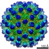
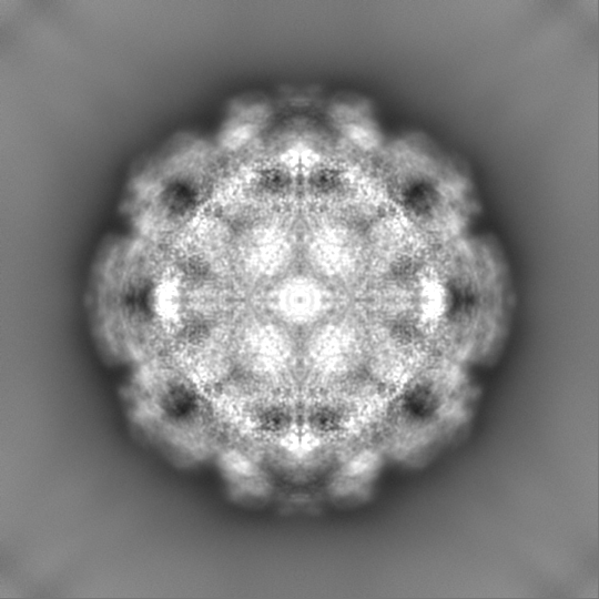
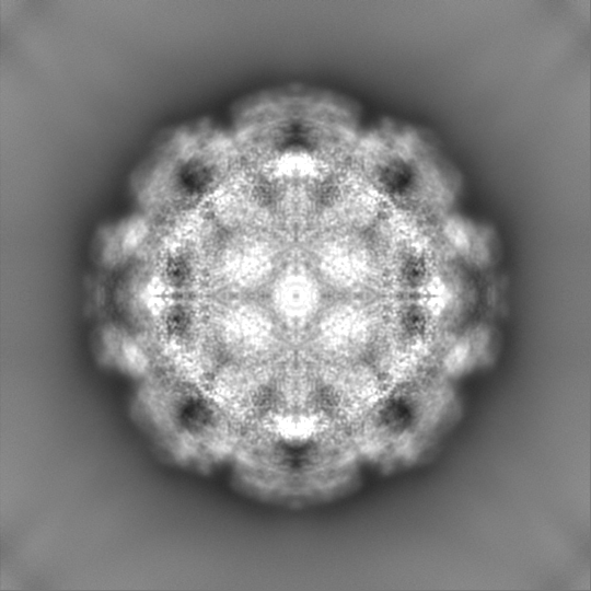
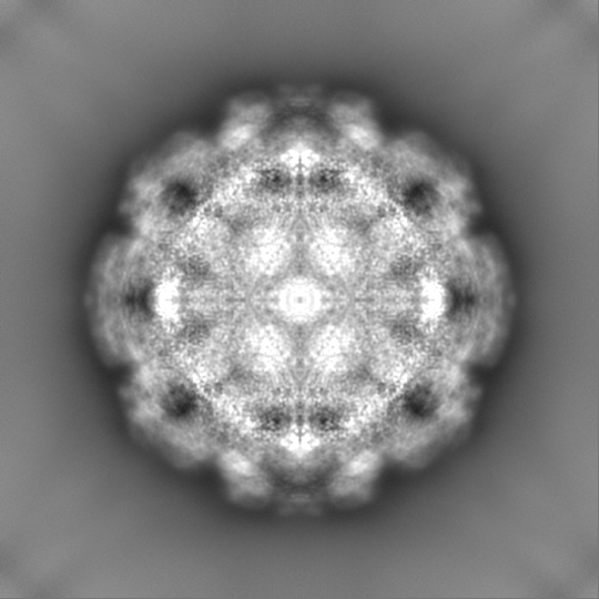
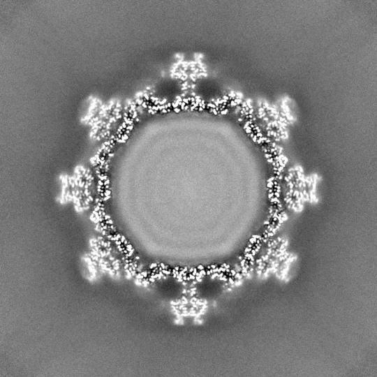
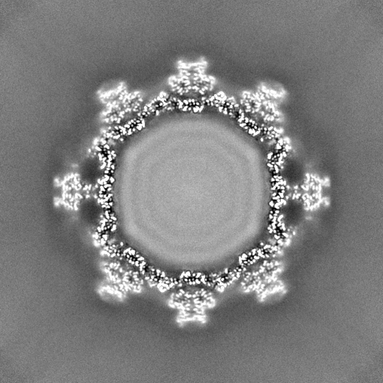
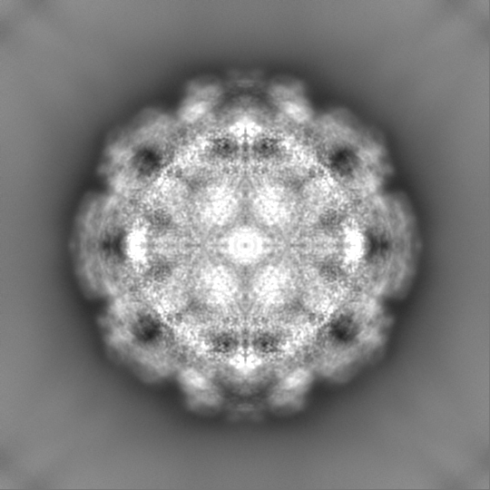
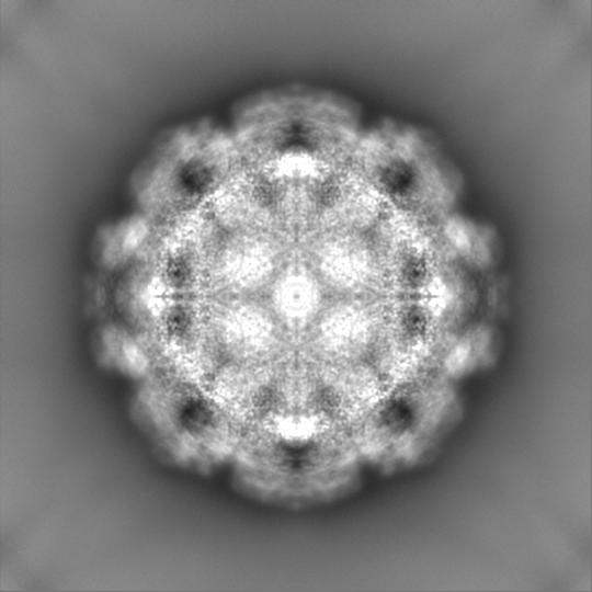
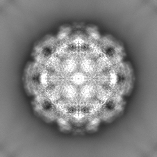
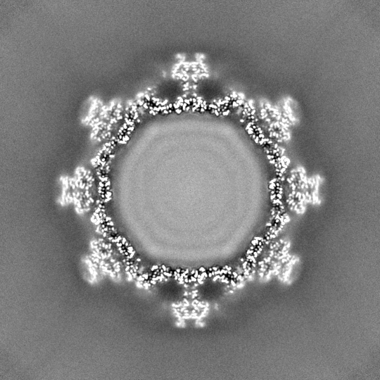
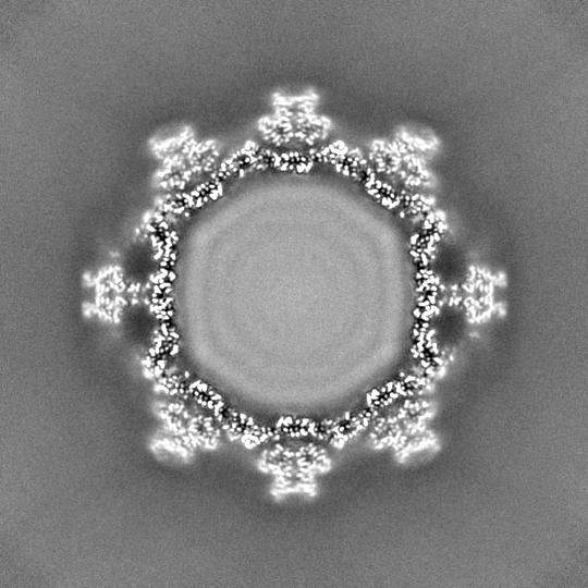
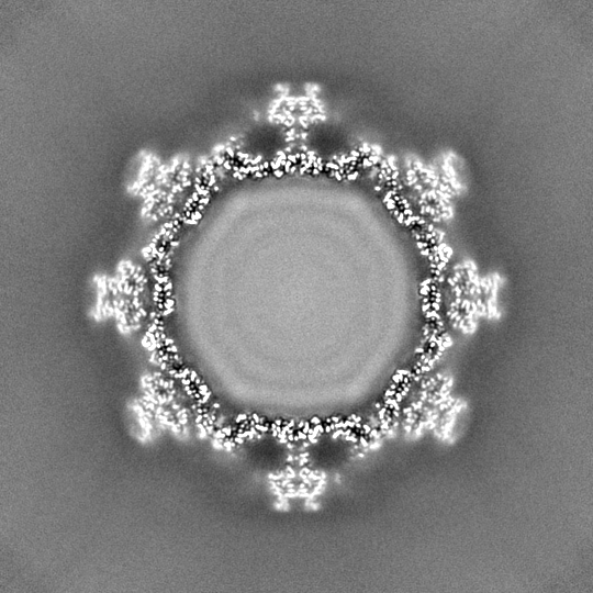
Journal: J Virol / Year: 2024 Title: The reversible activation of norovirus by metal ions. Authors: Michael Sherman / Faith Cox / Hong Smith / Mohamed H Habib / Stephanie Karst / Christiane E Wobus / Thomas J Smith / Abstract: Murine norovirus (MNV) undergoes extremely large conformational changes in response to the environment. The = 3 icosahedral capsid is composed of 180 copies of ~58-kDa VP1 comprised of N-terminus (N) ...Murine norovirus (MNV) undergoes extremely large conformational changes in response to the environment. The = 3 icosahedral capsid is composed of 180 copies of ~58-kDa VP1 comprised of N-terminus (N), shell (S), and C-terminal protruding (P) domains. At neutral pH, the P domains are loosely tethered to the shell and float ~15 Å above the surface. At low pH or in the presence of bile salts, the P domain drops onto the shell and this movement is accompanied by conformational changes within the P domain that enhance receptor interactions while blocking antibody binding. While previous crystallographic studies identified metal binding sites in the isolated P domain, the ~2.7-Å cryo-electron microscopy structures of MNV in the presence of Mg or Ca presented here show that metal ions can recapitulate the contraction observed at low pH or in the presence of bile. Further, we show that these conformational changes are reversed by dialysis against EDTA. As observed in the P domain crystal structures, metal ions bind to and contract the G'H' loop. This movement is correlated with the lifting of the C'D' loop and rotation of the P domain dimers about each other, exposing the bile salt binding pocket. Isothermal titration calorimetry experiments presented here demonstrate that the activation signals (bile salts, low pH, and metal ions) act in a synergistic manner that, individually, all result in the same activated structure. We present a model whereby these reversible conformational changes represent a uniquely dynamic and tissue-specific structural adaptation to the environment.IMPORTANCEThe highly mobile protruding domains on the calicivirus capsids are recognized by cell receptor(s) and antibodies. At neutral pH, they float ~15 Å above the shell but at low pH or in the presence of bile salts, they contract onto the surface. Concomitantly, changes within the P domain block antibody binding while enhancing receptor binding. While we previously demonstrated that metals also block antibody binding, it was unknown whether they might also cause similar conformational changes in the virion. Here, we present the near atomic cryo-electron microscopy structures of infectious murine norovirus (MNV) in the presence of calcium or magnesium ions. The metal ions reversibly induce the same P domain contraction as low pH and bile salts and act in a synergistic manner with the other stimuli. We propose that, unlike most other viruses, MNV facilely changes conformations as a unique means to escape immune surveillance as it moves through various tissues.
In the structure databanks used in Yorodumi, some data are registered as the other names, "COVID-19 virus" and "2019-nCoV". Here are the details of the virus and the list of structure data.
Jan 31, 2019. EMDB accession codes are about to change! (news from PDBe EMDB page)
EMDB accession codes are about to change! (news from PDBe EMDB page)
The allocation of 4 digits for EMDB accession codes will soon come to an end. Whilst these codes will remain in use, new EMDB accession codes will include an additional digit and will expand incrementally as the available range of codes is exhausted. The current 4-digit format prefixed with “EMD-” (i.e. EMD-XXXX) will advance to a 5-digit format (i.e. EMD-XXXXX), and so on. It is currently estimated that the 4-digit codes will be depleted around Spring 2019, at which point the 5-digit format will come into force.
The EM Navigator/Yorodumi systems omit the EMD- prefix.
Related info.:Q: What is EMD? / ID/Accession-code notation in Yorodumi/EM Navigator
Yorodumi is a browser for structure data from EMDB, PDB, SASBDB, etc.
This page is also the successor to EM Navigator detail page, and also detail information page/front-end page for Omokage search.
The word "yorodu" (or yorozu) is an old Japanese word meaning "ten thousand". "mi" (miru) is to see.
Related info.:EMDB / PDB / SASBDB / Comparison of 3 databanks / Yorodumi Search / Aug 31, 2016. New EM Navigator & Yorodumi / Yorodumi Papers / Jmol/JSmol / Function and homology information / Changes in new EM Navigator and Yorodumi
 Movie
Movie Controller


 Open data
 Basic information
 Map data
 Sample
 Keywords
 Function and homology information
 Murine norovirus 1
 Authors
 United States, 1 items
  Citation
 Structure visualization
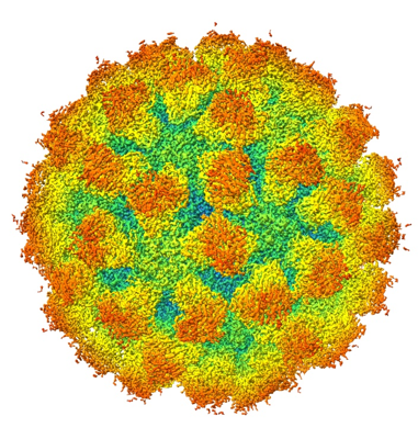
 Downloads & links
 emd_42604.png
 http://ftp.pdbj.org/pub/emdb/structures/EMD-42604


 Z (Sec.)
 Y (Row.)
 X (Col.)


 Sample components
 Processing
 Electron microscopy
 FIELD EMISSION GUN
