+ データを開く
- 基本情報
基本情報
| 登録情報 | データベース: PDB / ID: 8uk3 | ||||||
|---|---|---|---|---|---|---|---|
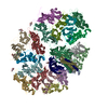
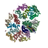
| タイトル | The rotavirus VP5*/VP8* conformational transition permeabilizes membranes to Ca2+ (class 6 reconstruction) | ||||||
 要素 要素 |
| ||||||
 キーワード キーワード | VIRAL PROTEIN / Non-enveloped virus / viral particle / entry / membrane-penetration / rotavirus / VP4 / VP5* / VP8* / calcium / Ca2+ / liposome | ||||||
| 機能・相同性 |  機能・相同性情報 機能・相同性情報host cell endoplasmic reticulum lumen / host cell rough endoplasmic reticulum / T=13 icosahedral viral capsid / permeabilization of host organelle membrane involved in viral entry into host cell / host cytoskeleton / viral outer capsid / host cell endoplasmic reticulum-Golgi intermediate compartment / receptor-mediated virion attachment to host cell / virion attachment to host cell / host cell plasma membrane ...host cell endoplasmic reticulum lumen / host cell rough endoplasmic reticulum / T=13 icosahedral viral capsid / permeabilization of host organelle membrane involved in viral entry into host cell / host cytoskeleton / viral outer capsid / host cell endoplasmic reticulum-Golgi intermediate compartment / receptor-mediated virion attachment to host cell / virion attachment to host cell / host cell plasma membrane / membrane / metal ion binding 類似検索 - 分子機能 | ||||||
| 生物種 |  Simian rotavirus A strain RRV (ウイルス) Simian rotavirus A strain RRV (ウイルス) | ||||||
| 手法 | 電子顕微鏡法 / 単粒子再構成法 / クライオ電子顕微鏡法 / 解像度: 8 Å | ||||||
 データ登録者 データ登録者 | De Sautu, M. / Herrmann, T. / Jenni, S. / Harrison, S.C. | ||||||
| 資金援助 |  米国, 1件 米国, 1件
| ||||||
 引用 引用 |  ジャーナル: PLoS Pathog / 年: 2024 ジャーナル: PLoS Pathog / 年: 2024タイトル: The rotavirus VP5*/VP8* conformational transition permeabilizes membranes to Ca2. 著者: Marilina de Sautu / Tobias Herrmann / Gustavo Scanavachi / Simon Jenni / Stephen C Harrison /  要旨: Rotaviruses infect cells by delivering into the cytosol a transcriptionally active inner capsid particle (a "double-layer particle": DLP). Delivery is the function of a third, outer layer, which ...Rotaviruses infect cells by delivering into the cytosol a transcriptionally active inner capsid particle (a "double-layer particle": DLP). Delivery is the function of a third, outer layer, which drives uptake from the cell surface into small vesicles from which the DLPs escape. In published work, we followed stages of rhesus rotavirus (RRV) entry by live-cell imaging and correlated them with structures from cryogenic electron microscopy and tomography (cryo-EM and cryo-ET). The virus appears to wrap itself in membrane, leading to complete engulfment and loss of Ca2+ from the vesicle produced by the wrapping. One of the outer-layer proteins, VP7, is a Ca2+-stabilized trimer; loss of Ca2+ releases both VP7 and the other outer-layer protein, VP4, from the particle. VP4, activated by cleavage into VP8* and VP5*, is a trimer that undergoes a large-scale conformational rearrangement, reminiscent of the transition that viral fusion proteins undergo to penetrate a membrane. The rearrangement of VP5* thrusts a 250-residue, C-terminal segment of each of the three subunits outward, while allowing the protein to remain attached to the virus particle and to the cell being infected. We proposed that this segment inserts into the membrane of the target cell, enabling Ca2+ to cross. In the work reported here, we show the validity of key aspects of this proposed sequence. By cryo-EM studies of liposome-attached virions ("triple-layer particles": TLPs) and single-particle fluorescence imaging of liposome-attached TLPs, we confirm insertion of the VP4 C-terminal segment into the membrane and ensuing generation of a Ca2+ "leak". The results allow us to formulate a molecular description of early events in entry. We also discuss our observations in the context of other work on double-strand RNA virus entry. | ||||||
| 履歴 |
|
- 構造の表示
構造の表示
| 構造ビューア | 分子:  Molmil Molmil Jmol/JSmol Jmol/JSmol |
|---|
- ダウンロードとリンク
- ダウンロード
| PDBx/mmCIF形式 |  8uk3.cif.gz 8uk3.cif.gz | 1.8 MB | 表示 |  PDBx/mmCIF形式 PDBx/mmCIF形式 |
|---|---|---|---|---|
| PDB形式 |  pdb8uk3.ent.gz pdb8uk3.ent.gz | 1.5 MB | 表示 |  PDB形式 PDB形式 |
| PDBx/mmJSON形式 |  8uk3.json.gz 8uk3.json.gz | ツリー表示 |  PDBx/mmJSON形式 PDBx/mmJSON形式 | |
| その他 |  その他のダウンロード その他のダウンロード |
-検証レポート
| 文書・要旨 |  8uk3_validation.pdf.gz 8uk3_validation.pdf.gz | 1.4 MB | 表示 |  wwPDB検証レポート wwPDB検証レポート |
|---|---|---|---|---|
| 文書・詳細版 |  8uk3_full_validation.pdf.gz 8uk3_full_validation.pdf.gz | 1.4 MB | 表示 | |
| XML形式データ |  8uk3_validation.xml.gz 8uk3_validation.xml.gz | 134.6 KB | 表示 | |
| CIF形式データ |  8uk3_validation.cif.gz 8uk3_validation.cif.gz | 210.6 KB | 表示 | |
| アーカイブディレクトリ |  https://data.pdbj.org/pub/pdb/validation_reports/uk/8uk3 https://data.pdbj.org/pub/pdb/validation_reports/uk/8uk3 ftp://data.pdbj.org/pub/pdb/validation_reports/uk/8uk3 ftp://data.pdbj.org/pub/pdb/validation_reports/uk/8uk3 | HTTPS FTP |
-関連構造データ
| 関連構造データ |  42344MC  8uk2C M: このデータのモデリングに利用したマップデータ C: 同じ文献を引用 ( |
|---|---|
| 類似構造データ | 類似検索 - 機能・相同性  F&H 検索 F&H 検索 |
- リンク
リンク
- 集合体
集合体
| 登録構造単位 | 
|
|---|---|
| 1 |
|
- 要素
要素
| #1: タンパク質 | 分子量: 86655.586 Da / 分子数: 3 / 由来タイプ: 組換発現 由来: (組換発現)  Simian rotavirus A strain RRV (ウイルス) Simian rotavirus A strain RRV (ウイルス)細胞株 (発現宿主): MA104 / 発現宿主:  Chlorocebus aethiops (ミドリザル) / 参照: UniProt: G0YZG6 Chlorocebus aethiops (ミドリザル) / 参照: UniProt: G0YZG6#2: タンパク質 | 分子量: 37136.531 Da / 分子数: 18 / 由来タイプ: 組換発現 由来: (組換発現)  Simian rotavirus A strain RRV (ウイルス) Simian rotavirus A strain RRV (ウイルス)細胞株 (発現宿主): MA104 / 発現宿主:  Chlorocebus aethiops (ミドリザル) / 参照: UniProt: P12476 Chlorocebus aethiops (ミドリザル) / 参照: UniProt: P12476#3: 糖 | ChemComp-NAG / #4: 化合物 | ChemComp-CA / 研究の焦点であるリガンドがあるか | N | Has protein modification | Y | |
|---|
-実験情報
-実験
| 実験 | 手法: 電子顕微鏡法 |
|---|---|
| EM実験 | 試料の集合状態: PARTICLE / 3次元再構成法: 単粒子再構成法 |
- 試料調製
試料調製
| 構成要素 | 名称: Simian rotavirus A strain RRV / タイプ: VIRUS / Entity ID: #1-#2 / 由来: NATURAL |
|---|---|
| 由来(天然) | 生物種:  Simian rotavirus A strain RRV (ウイルス) Simian rotavirus A strain RRV (ウイルス) |
| ウイルスについての詳細 | 中空か: NO / エンベロープを持つか: NO / 単離: STRAIN / タイプ: VIRION |
| 緩衝液 | pH: 8 |
| 試料 | 包埋: NO / シャドウイング: NO / 染色: NO / 凍結: YES |
| 急速凍結 | 凍結剤: ETHANE |
- 電子顕微鏡撮影
| 実験機器 |  モデル: Titan Krios / 画像提供: FEI Company |
|---|---|
| 顕微鏡 | モデル: FEI TITAN KRIOS |
| 電子銃 | 電子線源:  FIELD EMISSION GUN / 加速電圧: 300 kV / 照射モード: FLOOD BEAM FIELD EMISSION GUN / 加速電圧: 300 kV / 照射モード: FLOOD BEAM |
| 電子レンズ | モード: BRIGHT FIELD / 最大 デフォーカス(公称値): 2500 nm / 最小 デフォーカス(公称値): 1000 nm |
| 撮影 | 電子線照射量: 50 e/Å2 / フィルム・検出器のモデル: GATAN K3 (6k x 4k) |
- 解析
解析
| CTF補正 | タイプ: PHASE FLIPPING AND AMPLITUDE CORRECTION |
|---|---|
| 3次元再構成 | 解像度: 8 Å / 解像度の算出法: OTHER / 粒子像の数: 70018 / 対称性のタイプ: POINT |
| 原子モデル構築 | プロトコル: RIGID BODY FIT |
| 原子モデル構築 | PDB-ID: 6WXG Accession code: 6WXG / Source name: PDB / タイプ: experimental model |
 ムービー
ムービー コントローラー


 PDBj
PDBj


