 Movie
Movie Controller

[English] 日本語
 Yorodumi
- PDB-8u7m: Human retinal variant phosphomimetic IMPDH1(595)-S477D free octam... -
+ Open data

- Basic information
| Entry | Database: PDB / ID: 8u7m | ||||||||||||||||||
|---|---|---|---|---|---|---|---|---|---|---|---|---|---|---|---|---|---|---|---|
| Title | Human retinal variant phosphomimetic IMPDH1(595)-S477D free octamer bound by GTP, ATP, IMP, and NAD+ | ||||||||||||||||||
 Components Components | Inosine-5'-monophosphate dehydrogenase 1 | ||||||||||||||||||
 Keywords Keywords | OXIDOREDUCTASE / dehydrogenase / nucleotide synthesis / cytosol / phosphomimetic | ||||||||||||||||||
| Function / homology |  Function and homology information Function and homology informationPurine ribonucleoside monophosphate biosynthesis / IMP dehydrogenase activity / IMP dehydrogenase / GMP biosynthetic process / Azathioprine ADME / GTP biosynthetic process / azurophil granule lumen / secretory granule lumen / ficolin-1-rich granule lumen / Potential therapeutics for SARS ...Purine ribonucleoside monophosphate biosynthesis / IMP dehydrogenase activity / IMP dehydrogenase / GMP biosynthetic process / Azathioprine ADME / GTP biosynthetic process / azurophil granule lumen / secretory granule lumen / ficolin-1-rich granule lumen / Potential therapeutics for SARS / nucleic acid binding / nucleotide binding / Neutrophil degranulation / DNA binding / RNA binding / extracellular region / identical protein binding / nucleus / metal ion binding / cytosol / cytoplasm Similarity search - Function | ||||||||||||||||||
| Biological species |  Homo sapiens (human) Homo sapiens (human) | ||||||||||||||||||
| Method | ELECTRON MICROSCOPY / single particle reconstruction / cryo EM / Resolution: 3.1 Å | ||||||||||||||||||
 Authors Authors | Calise, S.J. / Kollman, J.M. | ||||||||||||||||||
| Funding support |  United States, 5items United States, 5items
| ||||||||||||||||||
 Citation Citation | Journal: J Cell Biol / Year: 2024 Title: Light-sensitive phosphorylation regulates retinal IMPDH1 activity and filament assembly. Authors: S John Calise / Audrey G O'Neill / Anika L Burrell / Miles S Dickinson / Josephine Molfino / Charlie Clarke / Joel Quispe / David Sokolov / Rubén M Buey / Justin M Kollman /  Abstract: Inosine monophosphate dehydrogenase (IMPDH) is the rate-limiting enzyme in guanosine triphosphate (GTP) synthesis and assembles into filaments in cells, which desensitizes the enzyme to feedback ...Inosine monophosphate dehydrogenase (IMPDH) is the rate-limiting enzyme in guanosine triphosphate (GTP) synthesis and assembles into filaments in cells, which desensitizes the enzyme to feedback inhibition and boosts nucleotide production. The vertebrate retina expresses two splice variants IMPDH1(546) and IMPDH1(595). In bovine retinas, residue S477 is preferentially phosphorylated in the dark, but the effects on IMPDH1 activity and regulation are unclear. Here, we generated phosphomimetic mutants to investigate structural and functional consequences of S477 phosphorylation. The S477D mutation resensitized both variants to GTP inhibition but only blocked assembly of IMPDH1(595) filaments. Cryo-EM structures of both variants showed that S477D specifically blocks assembly of a high-activity assembly interface, still allowing assembly of low-activity IMPDH1(546) filaments. Finally, we discovered that S477D exerts a dominant-negative effect in cells, preventing endogenous IMPDH filament assembly. By modulating the structure and higher-order assembly of IMPDH, S477 phosphorylation acts as a mechanism for downregulating retinal GTP synthesis in the dark when nucleotide turnover is decreased. | ||||||||||||||||||
| History |
|
- Structure visualization
| Structure viewer | Molecule: MolmilJmol/JSmol |
|---|
- Downloads & links
-Download
| PDBx/mmCIF format | 8u7m.cif.gz | 617.1 KB | Display | PDBx/mmCIF format |
|---|---|---|---|---|
| PDB format | pdb8u7m.ent.gz | 516 KB | Display | PDB format |
| PDBx/mmJSON format | 8u7m.json.gz | Tree view | PDBx/mmJSON format | |
| Others |  Other downloads Other downloads |
-Validation report
| Summary document | 8u7m_validation.pdf.gz | 3.5 MB | Display | wwPDB validaton report |
|---|---|---|---|---|
| Full document | 8u7m_full_validation.pdf.gz | 3.6 MB | Display | |
| Data in XML | 8u7m_validation.xml.gz | 117 KB | Display | |
| Data in CIF | 8u7m_validation.cif.gz | 162 KB | Display | |
| Arichive directory | https://data.pdbj.org/pub/pdb/validation_reports/u7/8u7mftp://data.pdbj.org/pub/pdb/validation_reports/u7/8u7m | HTTPS FTP |
-Related structure data
| Related structure data |  41986MC 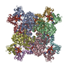 8u7qC 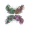 8u7vC 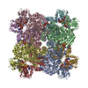 8u8oC 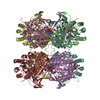 8u8yC M: map data used to model this data C: citing same article ( |
|---|---|
| Similar structure data |


-Links
 PDBj
PDBj
- Assembly
| Deposited unit | 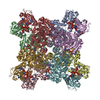
|
|---|---|
| 1 |
|
-Components
| #1: Protein | Mass: 63579.449 Da / Num. of mol.: 8 / Mutation: S477D Source method: isolated from a genetically manipulated source Source: (gene. exp.) Homo sapiens (human) / Gene: IMPDH1 / Production host:  #2: Chemical | ChemComp-GTP /   Mass: 523.180 Da / Num. of mol.: 16 / Source method: obtained synthetically / Formula: C10H16N5O14P3 / Feature type: SUBJECT OF INVESTIGATION / Comment: GTP, energy-carrying molecule*YM Mass: 523.180 Da / Num. of mol.: 16 / Source method: obtained synthetically / Formula: C10H16N5O14P3 / Feature type: SUBJECT OF INVESTIGATION / Comment: GTP, energy-carrying molecule*YM#3: Chemical | ChemComp-ATP /   Mass: 507.181 Da / Num. of mol.: 8 / Source method: obtained synthetically / Formula: C10H16N5O13P3 / Feature type: SUBJECT OF INVESTIGATION / Comment: ATP, energy-carrying molecule*YM Mass: 507.181 Da / Num. of mol.: 8 / Source method: obtained synthetically / Formula: C10H16N5O13P3 / Feature type: SUBJECT OF INVESTIGATION / Comment: ATP, energy-carrying molecule*YM#4: Chemical | ChemComp-IMP /   Mass: 348.206 Da / Num. of mol.: 8 / Source method: obtained synthetically / Formula: C10H13N4O8P / Feature type: SUBJECT OF INVESTIGATION Mass: 348.206 Da / Num. of mol.: 8 / Source method: obtained synthetically / Formula: C10H13N4O8P / Feature type: SUBJECT OF INVESTIGATION#5: Chemical | ChemComp-NAD /   Mass: 663.425 Da / Num. of mol.: 8 / Source method: obtained synthetically / Formula: C21H27N7O14P2 / Feature type: SUBJECT OF INVESTIGATION / Comment: NAD*YM Mass: 663.425 Da / Num. of mol.: 8 / Source method: obtained synthetically / Formula: C21H27N7O14P2 / Feature type: SUBJECT OF INVESTIGATION / Comment: NAD*YMHas ligand of interest | Y | |
|---|
-Experimental details
-Experiment
| Experiment | Method: ELECTRON MICROSCOPY |
|---|---|
| EM experiment | Aggregation state: PARTICLE / 3D reconstruction method: single particle reconstruction |
- Sample preparation
| Component | Name: IMPDH1(595)-S477D octamer bound by GTP, ATP, IMP, and NAD+ Type: COMPLEX / Entity ID: #1 / Source: RECOMBINANT |
|---|---|
| Molecular weight | Value: 0.508 MDa / Experimental value: NO |
| Source (natural) | Organism: Homo sapiens (human) |
| Source (recombinant) | Organism: |
| Buffer solution | pH: 7 |
| Specimen | Embedding applied: NO / Shadowing applied: NO / Staining applied: NO / Vitrification applied: YES |
| Specimen support | Grid material: COPPER / Grid type: C-flat-2/2 |
| Vitrification | Instrument: FEI VITROBOT MARK IV / Cryogen name: ETHANE |
- Electron microscopy imaging
| Microscopy | Model: TFS GLACIOS |
|---|---|
| Electron gun | Electron source:  FIELD EMISSION GUN / Accelerating voltage: 200 kV / Illumination mode: FLOOD BEAM FIELD EMISSION GUN / Accelerating voltage: 200 kV / Illumination mode: FLOOD BEAM |
| Electron lens | Mode: BRIGHT FIELD / Nominal magnification: 36000 X / Nominal defocus max: 1800 nm / Nominal defocus min: 1200 nm / Cs: 2.7 mm |
| Image recording | Electron dose: 65 e/Å2 / Detector mode: COUNTING / Film or detector model: GATAN K2 SUMMIT (4k x 4k) / Num. of real images: 646 |
- Processing
| EM software |
| ||||||||||||||||||||||||||||||||||||||||||||||||
|---|---|---|---|---|---|---|---|---|---|---|---|---|---|---|---|---|---|---|---|---|---|---|---|---|---|---|---|---|---|---|---|---|---|---|---|---|---|---|---|---|---|---|---|---|---|---|---|---|---|
| CTF correction | Type: PHASE FLIPPING AND AMPLITUDE CORRECTION | ||||||||||||||||||||||||||||||||||||||||||||||||
| Particle selection | Num. of particles selected: 343619 | ||||||||||||||||||||||||||||||||||||||||||||||||
| Symmetry | Point symmetry: D4 (2x4 fold dihedral) | ||||||||||||||||||||||||||||||||||||||||||||||||
| 3D reconstruction | Resolution: 3.1 Å / Resolution method: FSC 0.143 CUT-OFF / Num. of particles: 163814 / Symmetry type: POINT | ||||||||||||||||||||||||||||||||||||||||||||||||
| Refine LS restraints |
|
