 Movie
Movie Controller


+ Open data

- Basic information
| Entry | Database: PDB / ID: 8u62 | ||||||
|---|---|---|---|---|---|---|---|
| Title | Cryo-EM structure of PsBphP in Pfr state, Dimer of Dimers FL | ||||||
 Components Components | histidine kinase | ||||||
 Keywords Keywords | PLANT PROTEIN / Pseudomonas syringae bacteriophytochrome | ||||||
| Function / homology |  Function and homology information Function and homology informationdetection of visible light / phosphorelay sensor kinase activity / histidine kinase / photoreceptor activity / regulation of DNA-templated transcription / ATP binding / plasma membrane Similarity search - Function | ||||||
| Biological species |  Pseudomonas syringae pv. tomato str. DC3000 (bacteria) Pseudomonas syringae pv. tomato str. DC3000 (bacteria) | ||||||
| Method | ELECTRON MICROSCOPY / single particle reconstruction / cryo EM / Resolution: 3.3 Å | ||||||
 Authors Authors | Basore, K. / Burgie, E.S. / Vierstra, D. | ||||||
| Funding support |  United States, 1items United States, 1items
| ||||||
 Citation Citation | Journal: Nat Commun / Year: 2024 Title: Signaling by a bacterial phytochrome histidine kinase involves a conformational cascade reorganizing the dimeric photoreceptor. Authors: E Sethe Burgie / Katherine Basore / Michael J Rau / Brock Summers / Alayna J Mickles / Vadim Grigura / James A J Fitzpatrick / Richard D Vierstra /  Abstract: Phytochromes (Phys) are a divergent cohort of bili-proteins that detect light through reversible interconversion between dark-adapted Pr and photoactivated Pfr states. While our understandings of ...Phytochromes (Phys) are a divergent cohort of bili-proteins that detect light through reversible interconversion between dark-adapted Pr and photoactivated Pfr states. While our understandings of downstream events are emerging, it remains unclear how Phys translate light into an interpretable conformational signal. Here, we present models of both states for a dimeric Phy with histidine kinase (HK) activity from the proteobacterium Pseudomonas syringae, which were built from high-resolution cryo-EM maps (2.8-3.4-Å) of the photosensory module (PSM) and its following signaling (S) helix together with lower resolution maps for the downstream output region augmented by RoseTTAFold and AlphaFold structural predictions. The head-to-head models reveal the PSM and its photointerconversion mechanism with strong clarity, while the HK region is interpretable but relatively mobile. Pr/Pfr comparisons show that bilin phototransformation alters PSM architecture culminating in a scissoring motion of the paired S-helices linking the PSMs to the HK bidomains that ends in reorientation of the paired catalytic ATPase modules relative to the phosphoacceptor histidines. This action apparently primes autophosphorylation enroute to phosphotransfer to the cognate DNA-binding response regulator AlgB which drives quorum-sensing behavior through transient association with the photoreceptor. Collectively, these models illustrate how light absorption conformationally translates into accelerated signaling by Phy-type kinases. | ||||||
| History |
|
- Structure visualization
| Structure viewer | Molecule: MolmilJmol/JSmol |
|---|
- Downloads & links
-Download
| PDBx/mmCIF format | 8u62.cif.gz | 211.1 KB | Display | PDBx/mmCIF format |
|---|---|---|---|---|
| PDB format | pdb8u62.ent.gz | 163.7 KB | Display | PDB format |
| PDBx/mmJSON format | 8u62.json.gz | Tree view | PDBx/mmJSON format | |
| Others |  Other downloads Other downloads |
-Validation report
| Arichive directory | https://data.pdbj.org/pub/pdb/validation_reports/u6/8u62ftp://data.pdbj.org/pub/pdb/validation_reports/u6/8u62 | HTTPS FTP |
|---|
-Related structure data
| Related structure data | 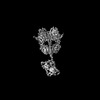 41941MC 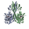 8u4xC 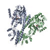 8u63C 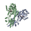 8u64C 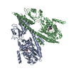 8u65C 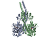 8u8zC M: map data used to model this data C: citing same article ( |
|---|---|
| Similar structure data |


-Links
 PDBj
PDBj


- Assembly
| Deposited unit | 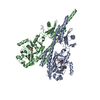
|
|---|---|
| 1 |
|
-Components
| #1: Protein | Mass: 82382.555 Da / Num. of mol.: 2 Source method: isolated from a genetically manipulated source Source: (gene. exp.) Pseudomonas syringae pv. tomato str. DC3000 (bacteria)Gene: bphP Plasmid: pBADCDS includes N-terminal TEV protease cleavable hexahistidine tag Production host: #2: Chemical | 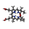  Mass: 585.670 Da / Num. of mol.: 2 / Source method: obtained synthetically / Formula: C33H37N4O6 / Feature type: SUBJECT OF INVESTIGATION Mass: 585.670 Da / Num. of mol.: 2 / Source method: obtained synthetically / Formula: C33H37N4O6 / Feature type: SUBJECT OF INVESTIGATIONHas ligand of interest | Y | Has protein modification | Y | |
|---|
-Experimental details
-Experiment
| Experiment | Method: ELECTRON MICROSCOPY |
|---|---|
| EM experiment | Aggregation state: PARTICLE / 3D reconstruction method: single particle reconstruction |
- Sample preparation
| Component | Name: PsBphP / Type: COMPLEX / Entity ID: #1 / Source: RECOMBINANT |
|---|---|
| Molecular weight | Experimental value: NO |
| Source (natural) | Organism: Pseudomonas syringae pv. tomato str. DC3000 (bacteria) |
| Source (recombinant) | Organism: |
| Buffer solution | pH: 7.5 |
| Specimen | Conc.: 0.5 mg/ml / Embedding applied: NO / Shadowing applied: NO / Staining applied: NO / Vitrification applied: YES |
| Vitrification | Instrument: FEI VITROBOT MARK IV / Cryogen name: ETHANE / Humidity: 100 % / Chamber temperature: 277 K |
- Electron microscopy imaging
| Experimental equipment |  Model: Titan Krios / Image courtesy: FEI Company |
|---|---|
| Microscopy | Model: FEI TITAN KRIOS / Details: Preliminary grid screening was performed manually. |
| Electron gun | Electron source:  FIELD EMISSION GUN / Accelerating voltage: 300 kV / Illumination mode: FLOOD BEAM FIELD EMISSION GUN / Accelerating voltage: 300 kV / Illumination mode: FLOOD BEAM |
| Electron lens | Mode: BRIGHT FIELD / Nominal magnification: 96000 X / Nominal defocus max: 2400 nm / Nominal defocus min: 1000 nm / Cs: 0.01 mm / C2 aperture diameter: 150 µm / Alignment procedure: ZEMLIN TABLEAU |
| Specimen holder | Cryogen: NITROGEN / Specimen holder model: FEI TITAN KRIOS AUTOGRID HOLDER / Temperature (max): 84 K / Temperature (min): 82 K |
| Image recording | Average exposure time: 4.28 sec. / Electron dose: 51.78 e/Å2 / Film or detector model: FEI FALCON IV (4k x 4k) / Num. of grids imaged: 3 / Num. of real images: 12593 |
| EM imaging optics | Spherical aberration corrector: Microscope is outfitted with a Cs image corrector with two hexapole elements. |
| Image scans | Width: 4096 / Height: 4096 |
- Processing
| EM software |
| ||||||||||||||||||||||||||||||||||||||||
|---|---|---|---|---|---|---|---|---|---|---|---|---|---|---|---|---|---|---|---|---|---|---|---|---|---|---|---|---|---|---|---|---|---|---|---|---|---|---|---|---|---|
| CTF correction | Type: PHASE FLIPPING AND AMPLITUDE CORRECTION | ||||||||||||||||||||||||||||||||||||||||
| Particle selection | Num. of particles selected: 4796622 | ||||||||||||||||||||||||||||||||||||||||
| 3D reconstruction | Resolution: 3.3 Å / Resolution method: FSC 0.143 CUT-OFF / Num. of particles: 402598 / Symmetry type: POINT | ||||||||||||||||||||||||||||||||||||||||
| Refine LS restraints |
|
