National Institutes of Health/National Institute of General Medical Sciences (NIH/NIGMS)
R01 GM127892
United States
Citation
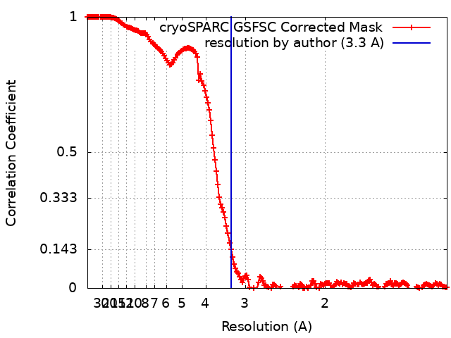
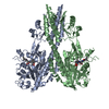
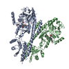
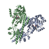
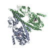
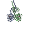
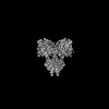
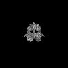
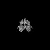
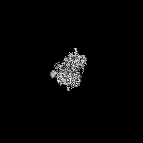
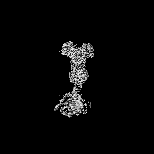
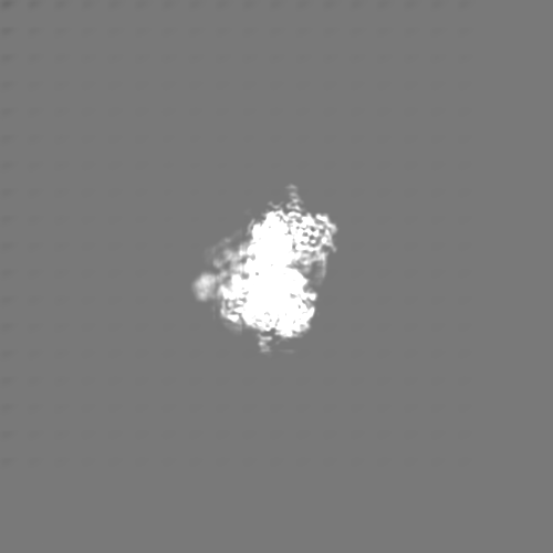
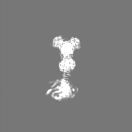
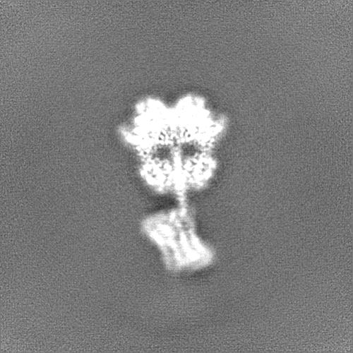
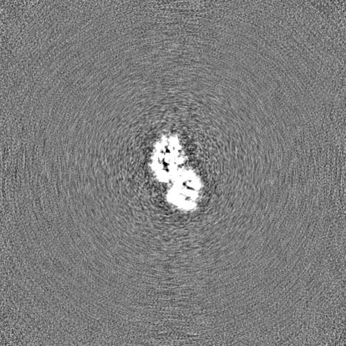
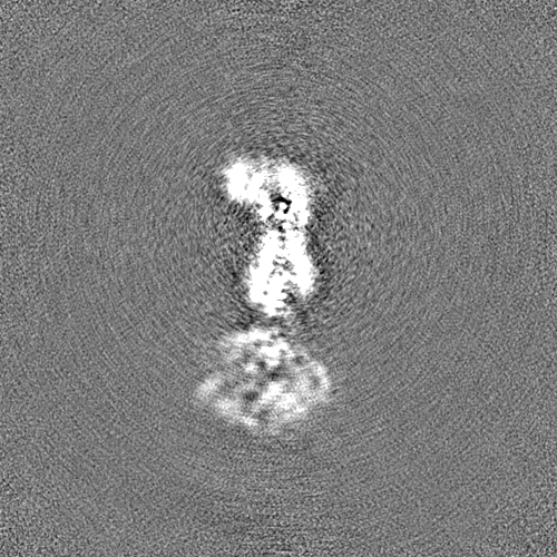
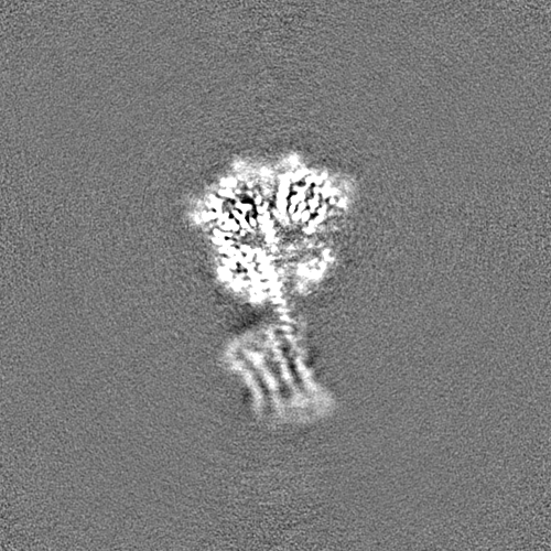
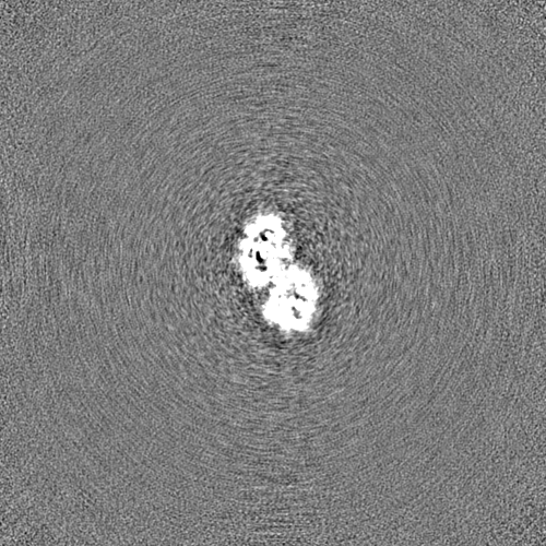
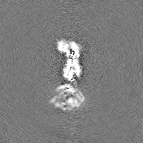
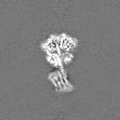
Journal: Nat Commun / Year: 2024 Title: Signaling by a bacterial phytochrome histidine kinase involves a conformational cascade reorganizing the dimeric photoreceptor. Authors: E Sethe Burgie / Katherine Basore / Michael J Rau / Brock Summers / Alayna J Mickles / Vadim Grigura / James A J Fitzpatrick / Richard D Vierstra / Abstract: Phytochromes (Phys) are a divergent cohort of bili-proteins that detect light through reversible interconversion between dark-adapted Pr and photoactivated Pfr states. While our understandings of ...Phytochromes (Phys) are a divergent cohort of bili-proteins that detect light through reversible interconversion between dark-adapted Pr and photoactivated Pfr states. While our understandings of downstream events are emerging, it remains unclear how Phys translate light into an interpretable conformational signal. Here, we present models of both states for a dimeric Phy with histidine kinase (HK) activity from the proteobacterium Pseudomonas syringae, which were built from high-resolution cryo-EM maps (2.8-3.4-Å) of the photosensory module (PSM) and its following signaling (S) helix together with lower resolution maps for the downstream output region augmented by RoseTTAFold and AlphaFold structural predictions. The head-to-head models reveal the PSM and its photointerconversion mechanism with strong clarity, while the HK region is interpretable but relatively mobile. Pr/Pfr comparisons show that bilin phototransformation alters PSM architecture culminating in a scissoring motion of the paired S-helices linking the PSMs to the HK bidomains that ends in reorientation of the paired catalytic ATPase modules relative to the phosphoacceptor histidines. This action apparently primes autophosphorylation enroute to phosphotransfer to the cognate DNA-binding response regulator AlgB which drives quorum-sensing behavior through transient association with the photoreceptor. Collectively, these models illustrate how light absorption conformationally translates into accelerated signaling by Phy-type kinases.
Cryogen name: ETHANE / Chamber humidity: 100 % / Chamber temperature: 277 K / Instrument: FEI VITROBOT MARK IV
-
Electron microscopy
Microscope
FEI TITAN KRIOS
Temperature
Min: 82.0 K / Max: 84.0 K
Specialist optics
Spherical aberration corrector: Microscope is outfitted with a Cs image corrector with two hexapole elements.
Details
Preliminary grid screening was performed manually.
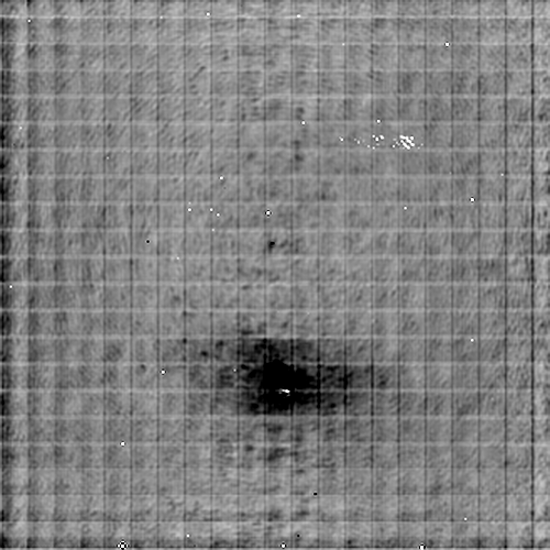
Image recording
Film or detector model: FEI FALCON IV (4k x 4k) / Digitization - Dimensions - Width: 4096 pixel / Digitization - Dimensions - Height: 4096 pixel / Number grids imaged: 3 / Number real images: 12593 / Average exposure time: 4.28 sec. / Average electron dose: 51.78 e/Å2
Electron beam
Acceleration voltage: 300 kV / Electron source: FIELD EMISSION GUN
In the structure databanks used in Yorodumi, some data are registered as the other names, "COVID-19 virus" and "2019-nCoV". Here are the details of the virus and the list of structure data.
Jan 31, 2019. EMDB accession codes are about to change! (news from PDBe EMDB page)
EMDB accession codes are about to change! (news from PDBe EMDB page)
The allocation of 4 digits for EMDB accession codes will soon come to an end. Whilst these codes will remain in use, new EMDB accession codes will include an additional digit and will expand incrementally as the available range of codes is exhausted. The current 4-digit format prefixed with “EMD-” (i.e. EMD-XXXX) will advance to a 5-digit format (i.e. EMD-XXXXX), and so on. It is currently estimated that the 4-digit codes will be depleted around Spring 2019, at which point the 5-digit format will come into force.
The EM Navigator/Yorodumi systems omit the EMD- prefix.
Related info.:Q: What is EMD? / ID/Accession-code notation in Yorodumi/EM Navigator
Yorodumi is a browser for structure data from EMDB, PDB, SASBDB, etc.
This page is also the successor to EM Navigator detail page, and also detail information page/front-end page for Omokage search.
The word "yorodu" (or yorozu) is an old Japanese word meaning "ten thousand". "mi" (miru) is to see.
Related info.:EMDB / PDB / SASBDB / Comparison of 3 databanks / Yorodumi Search / Aug 31, 2016. New EM Navigator & Yorodumi / Yorodumi Papers / Jmol/JSmol / Function and homology information / Changes in new EM Navigator and Yorodumi
 Movie
Movie Controller


 Open data
 Basic information
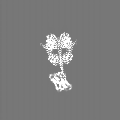
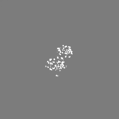
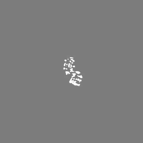
 Map data
 Sample
 Keywords
 Function and homology information
 Pseudomonas syringae pv. tomato str. DC3000 (bacteria)
 Authors
 United States, 1 items
  Citation
 Structure visualization
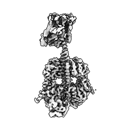
 Downloads & links
 emd_41941.png
 http://ftp.pdbj.org/pub/emdb/structures/EMD-41941


 Z (Sec.)
 Y (Row.)
 X (Col.)


 Sample components
 Processing
 Electron microscopy
 FIELD EMISSION GUN