 Movie
Movie Controller

[English] 日本語
 Yorodumi
- PDB-8u5h: Cryo-EM structure of human DNMT3A UDR bound to H2AK119ub1-modifie... -
+ Open data

- Basic information
| Entry | Database: PDB / ID: 8u5h | |||||||||||||||||||||||||||
|---|---|---|---|---|---|---|---|---|---|---|---|---|---|---|---|---|---|---|---|---|---|---|---|---|---|---|---|---|
| Title | Cryo-EM structure of human DNMT3A UDR bound to H2AK119ub1-modified nucleosome | |||||||||||||||||||||||||||
 Components Components |
| |||||||||||||||||||||||||||
 Keywords Keywords | STRUCTURAL PROTEIN/TRANSFERASE/DNA / DNA cytosine methyltransferase / STRUCTURAL PROTEIN-TRANSFERASE-DNA complex | |||||||||||||||||||||||||||
| Function / homology |  Function and homology information Function and homology informationtransposable element silencing by piRNA-mediated DNA methylation / protein-cysteine methyltransferase activity / positive regulation of cellular response to hypoxia / regulatory ncRNA-mediated heterochromatin formation / cellular response to bisphenol A / unmethylated CpG binding / DNA (cytosine-5-)-methyltransferase / DNA (cytosine-5-)-methyltransferase activity / autosome genomic imprinting / SUMOylation of DNA methylation proteins ...transposable element silencing by piRNA-mediated DNA methylation / protein-cysteine methyltransferase activity / positive regulation of cellular response to hypoxia / regulatory ncRNA-mediated heterochromatin formation / cellular response to bisphenol A / unmethylated CpG binding / DNA (cytosine-5-)-methyltransferase / DNA (cytosine-5-)-methyltransferase activity / autosome genomic imprinting / SUMOylation of DNA methylation proteins / XY body / oocyte development / response to vitamin A / response to ionizing radiation / DNA methylation-dependent constitutive heterochromatin formation / hepatocyte apoptotic process / negative regulation of gene expression via chromosomal CpG island methylation / lncRNA binding / chromosome, centromeric region / cellular response to ethanol / catalytic complex / heterochromatin / Maturation of protein E / Maturation of protein E / ER Quality Control Compartment (ERQC) / Myoclonic epilepsy of Lafora / FLT3 signaling by CBL mutants / IRAK2 mediated activation of TAK1 complex / Alpha-protein kinase 1 signaling pathway / Glycogen synthesis / IRAK1 recruits IKK complex / IRAK1 recruits IKK complex upon TLR7/8 or 9 stimulation / Prevention of phagosomal-lysosomal fusion / Endosomal Sorting Complex Required For Transport (ESCRT) / Membrane binding and targetting of GAG proteins / Negative regulation of FLT3 / Regulation of TBK1, IKKε (IKBKE)-mediated activation of IRF3, IRF7 / PTK6 Regulates RTKs and Their Effectors AKT1 and DOK1 / Regulation of TBK1, IKKε-mediated activation of IRF3, IRF7 upon TLR3 ligation / IRAK2 mediated activation of TAK1 complex upon TLR7/8 or 9 stimulation / Constitutive Signaling by NOTCH1 HD Domain Mutants / NOTCH2 Activation and Transmission of Signal to the Nucleus / TICAM1,TRAF6-dependent induction of TAK1 complex / TICAM1-dependent activation of IRF3/IRF7 / APC/C:Cdc20 mediated degradation of Cyclin B / Downregulation of ERBB4 signaling / Regulation of FZD by ubiquitination / APC-Cdc20 mediated degradation of Nek2A / Transferases; Transferring one-carbon groups; Methyltransferases / p75NTR recruits signalling complexes / InlA-mediated entry of Listeria monocytogenes into host cells / TRAF6 mediated IRF7 activation in TLR7/8 or 9 signaling / NF-kB is activated and signals survival / TRAF6-mediated induction of TAK1 complex within TLR4 complex / Regulation of pyruvate metabolism / Pexophagy / Downregulation of ERBB2:ERBB3 signaling / Regulation of innate immune responses to cytosolic DNA / NRIF signals cell death from the nucleus / Regulation of PTEN localization / VLDLR internalisation and degradation / DNA methylation / Activated NOTCH1 Transmits Signal to the Nucleus / Synthesis of active ubiquitin: roles of E1 and E2 enzymes / Translesion synthesis by REV1 / TICAM1, RIP1-mediated IKK complex recruitment / Regulation of BACH1 activity / Translesion synthesis by POLK / InlB-mediated entry of Listeria monocytogenes into host cell / JNK (c-Jun kinases) phosphorylation and activation mediated by activated human TAK1 / MAP3K8 (TPL2)-dependent MAPK1/3 activation / Activation of IRF3, IRF7 mediated by TBK1, IKKε (IKBKE) / Downregulation of TGF-beta receptor signaling / Translesion synthesis by POLI / post-embryonic development / PRC2 methylates histones and DNA / Josephin domain DUBs / Gap-filling DNA repair synthesis and ligation in GG-NER / IKK complex recruitment mediated by RIP1 / PINK1-PRKN Mediated Mitophagy / TGF-beta receptor signaling in EMT (epithelial to mesenchymal transition) / Regulation of activated PAK-2p34 by proteasome mediated degradation / Defective pyroptosis / TNFR1-induced NF-kappa-B signaling pathway / TCF dependent signaling in response to WNT / Regulation of NF-kappa B signaling / activated TAK1 mediates p38 MAPK activation / response to cocaine / Autodegradation of Cdh1 by Cdh1:APC/C / APC/C:Cdc20 mediated degradation of Securin / Regulation of endogenous retroelements by Piwi-interacting RNAs (piRNAs) / N-glycan trimming in the ER and Calnexin/Calreticulin cycle / Asymmetric localization of PCP proteins / cellular response to amino acid stimulus / Ubiquitin-dependent degradation of Cyclin D / NOTCH3 Activation and Transmission of Signal to the Nucleus / Regulation of signaling by CBL / Negative regulators of DDX58/IFIH1 signaling / Fanconi Anemia Pathway / Negative regulation of FGFR3 signaling Similarity search - Function | |||||||||||||||||||||||||||
| Biological species |  Homo sapiens (human) Homo sapiens (human) | |||||||||||||||||||||||||||
| Method | ELECTRON MICROSCOPY / single particle reconstruction / cryo EM / Resolution: 3.23 Å | |||||||||||||||||||||||||||
 Authors Authors | Chen, X. / Song, J. | |||||||||||||||||||||||||||
| Funding support |  United States, 1items United States, 1items
| |||||||||||||||||||||||||||
 Citation Citation | Journal: Nat Commun / Year: 2024 Title: Structural basis for the H2AK119ub1-specific DNMT3A-nucleosome interaction. Authors: Xinyi Chen / Yiran Guo / Ting Zhao / Jiuwei Lu / Jian Fang / Yinsheng Wang / Gang Greg Wang / Jikui Song / Abstract: Isoform 1 of DNA methyltransferase DNMT3A (DNMT3A1) specifically recognizes nucleosome monoubiquitylated at histone H2A lysine-119 (H2AK119ub1) for establishment of DNA methylation. Mis-regulation of ...Isoform 1 of DNA methyltransferase DNMT3A (DNMT3A1) specifically recognizes nucleosome monoubiquitylated at histone H2A lysine-119 (H2AK119ub1) for establishment of DNA methylation. Mis-regulation of this process may cause aberrant DNA methylation and pathogenesis. However, the molecular basis underlying DNMT3A1-nucleosome interaction remains elusive. Here we report the cryo-EM structure of DNMT3A1's ubiquitin-dependent recruitment (UDR) fragment complexed with H2AK119ub1-modified nucleosome. DNMT3A1 UDR occupies an extensive nucleosome surface, involving the H2A-H2B acidic patch, a surface groove formed by H2A and H3, nucleosomal DNA, and H2AK119ub1. The DNMT3A1 UDR's interaction with H2AK119ub1 affects the functionality of DNMT3A1 in cells in a context-dependent manner. Our structural and biochemical analysis also reveals competition between DNMT3A1 and JARID2, a cofactor of polycomb repression complex 2 (PRC2), for nucleosome binding, suggesting the interplay between different epigenetic pathways. Together, this study reports a molecular basis for H2AK119ub1-dependent DNMT3A1-nucleosome association, with important implications in DNMT3A1-mediated DNA methylation in development. | |||||||||||||||||||||||||||
| History |
|
- Structure visualization
| Structure viewer | Molecule: MolmilJmol/JSmol |
|---|
- Downloads & links
-Download
| PDBx/mmCIF format | 8u5h.cif.gz | 361.3 KB | Display | PDBx/mmCIF format |
|---|---|---|---|---|
| PDB format | pdb8u5h.ent.gz | 270.5 KB | Display | PDB format |
| PDBx/mmJSON format | 8u5h.json.gz | Tree view | PDBx/mmJSON format | |
| Others |  Other downloads Other downloads |
-Validation report
| Arichive directory | https://data.pdbj.org/pub/pdb/validation_reports/u5/8u5hftp://data.pdbj.org/pub/pdb/validation_reports/u5/8u5h | HTTPS FTP |
|---|
-Related structure data
| Related structure data |  41922MC M: map data used to model this data C: citing same article ( |
|---|---|
| Similar structure data |


-Links
 PDBj
PDBj


- Assembly
| Deposited unit | 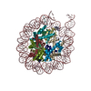
|
|---|---|
| 1 |
|
-Components
-Protein , 6 types, 11 molecules ABDIOJQKRMS
| #1: Protein | Mass: 11070.631 Da / Num. of mol.: 2 Source method: isolated from a genetically manipulated source Source: (gene. exp.) Homo sapiens (human) / Gene: DNMT3A / Production host:  #3: Protein | | Mass: 8576.831 Da / Num. of mol.: 1 Source method: isolated from a genetically manipulated source Source: (gene. exp.) Homo sapiens (human) / Gene: UBC / Production host: #5: Protein | Mass: 15521.349 Da / Num. of mol.: 2 Source method: isolated from a genetically manipulated source Source: (gene. exp.) #6: Protein | Mass: 11394.426 Da / Num. of mol.: 2 Source method: isolated from a genetically manipulated source Source: (gene. exp.) #7: Protein | Mass: 14083.398 Da / Num. of mol.: 2 Source method: isolated from a genetically manipulated source Source: (gene. exp.) #8: Protein | Mass: 13655.948 Da / Num. of mol.: 2 Source method: isolated from a genetically manipulated source Source: (gene. exp.) |
|---|
-DNA chain , 2 types, 2 molecules CH
| #2: DNA chain | Mass: 55176.137 Da / Num. of mol.: 1 Source method: isolated from a genetically manipulated source Source: (gene. exp.) Homo sapiens (human) / Production host: |
|---|---|
| #4: DNA chain | Mass: 55963.656 Da / Num. of mol.: 1 Source method: isolated from a genetically manipulated source Source: (gene. exp.) Homo sapiens (human) / Production host: |
-Details
| Has protein modification | N |
|---|
-Experimental details
-Experiment
| Experiment | Method: ELECTRON MICROSCOPY |
|---|---|
| EM experiment | Aggregation state: PARTICLE / 3D reconstruction method: single particle reconstruction |
- Sample preparation
| Component | Name: DNMT3A N-terminal domain and H2AK119Ub nucleosome complex Type: COMPLEX / Entity ID: all / Source: MULTIPLE SOURCES |
|---|---|
| Source (natural) | Organism: Homo sapiens (human) |
| Source (recombinant) | Organism: |
| Buffer solution | pH: 7.5 |
| Specimen | Embedding applied: NO / Shadowing applied: NO / Staining applied: NO / Vitrification applied: YES |
| Vitrification | Cryogen name: ETHANE |
- Electron microscopy imaging
| Experimental equipment |  Model: Titan Krios / Image courtesy: FEI Company |
|---|---|
| Microscopy | Model: FEI TITAN KRIOS |
| Electron gun | Electron source:  FIELD EMISSION GUN / Accelerating voltage: 300 kV / Illumination mode: FLOOD BEAM FIELD EMISSION GUN / Accelerating voltage: 300 kV / Illumination mode: FLOOD BEAM |
| Electron lens | Mode: BRIGHT FIELD / Nominal defocus max: 2500 nm / Nominal defocus min: 800 nm |
| Image recording | Electron dose: 50 e/Å2 / Film or detector model: FEI FALCON IV (4k x 4k) |
- Processing
| EM software | Name: PHENIX / Category: model refinement | ||||||||||||||||||||||||
|---|---|---|---|---|---|---|---|---|---|---|---|---|---|---|---|---|---|---|---|---|---|---|---|---|---|
| CTF correction | Type: PHASE FLIPPING AND AMPLITUDE CORRECTION | ||||||||||||||||||||||||
| 3D reconstruction | Resolution: 3.23 Å / Resolution method: FSC 0.143 CUT-OFF / Num. of particles: 86685 / Symmetry type: POINT | ||||||||||||||||||||||||
| Refine LS restraints |
|
